
How Soon Can You See a Heartbeat on Ultrasound? 2026
Cardiac activity can often be seen on a transvaginal ultrasound at about 6 weeks of pregnancy, counted from the first day of your last menstrual period. An abdominal scan usually shows it later, around 7 to 8 weeks. If a scan shows no cardiac activity earlier than expected, the result needs to be interpreted alongside the pregnancy measurements and your clinical history; it does not, by itself, establish a pregnancy loss.
TL;DR
A transvaginal ultrasound can often show cardiac activity at about 6 weeks; an abdominal scan usually shows it around 7 to 8 weeks.
For the question of how soon can you see a heartbeat on ultrasound, scan type and pregnancy dating change the answer.
The Ultrasound Concierge is best for Keller and Willow Park patients seeking a private pregnancy ultrasound, not urgent assessment.
No visible cardiac activity on a very early scan is not, by itself, a diagnosis of pregnancy loss.
How soon can you see a heartbeat on ultrasound?
About 6 weeks is the earliest common answer for a transvaginal scan; around 7 to 8 weeks is a more useful expectation for an abdominal scan. These are guides, not deadlines. In 2026, as in any pregnancy, what the sonographer can see depends on how far the pregnancy has developed and which structures are visible, not just the date on your calendar.
Transvaginal scan
When cardiac activity is commonly visible: About 6 weeks
What a result tells you: Offers the clearest early view when the pregnancy is still small.
Abdominal scan
When cardiac activity is commonly visible: Often around 7 to 8 weeks
What a result tells you: An early inconclusive view can reflect the limits of this approach.
Follow-up scan
When cardiac activity is commonly visible: Timing set by your clinician
What a result tells you: Shows whether expected development has occurred since the earlier scan.
The term heartbeat is familiar, but on an early scan the clinician is looking for cardiac activity: visible motion in the developing embryo. Seeing that motion is different from hearing a heartbeat. You do not need an audio recording for a clinician to identify cardiac activity on ultrasound.
Why this matters
An early scan can answer different questions: whether a pregnancy is visible in the uterus, whether an embryo can be identified, and whether cardiac activity can be seen. Those answers do not always arrive on the same visit. If you are considering a private appointment with The Ultrasound Concierge in the Keller or Willow Park area, tell the provider how your pregnancy was dated and whether you need a diagnostic assessment or an elective scan.
An inconclusive early scan is a finding to follow up, not a verdict to interpret alone. The right next step depends on the images, measurements, symptoms and your clinician’s assessment. This distinction matters most when you expected to see cardiac activity and did not.
Transvaginal ultrasound: about 6 weeks
A transvaginal scan places the ultrasound probe in the vagina, closer to the uterus. That closer view makes it the more informative approach when a pregnancy is too early to assess clearly through the abdomen. Cardiac activity can often be seen at about 6 weeks, but an individual scan can still be inconclusive at that point.
Pregnancy weeks are usually counted from the first day of the last menstrual period, not from conception. If ovulation happened later than expected, a pregnancy described as 6 weeks by period dates can be less developed on ultrasound. A clinician uses the scan findings and dating information together rather than treating the calendar estimate as a measurement of the embryo.
If the scan shows a gestational sac but no embryo, the question is not simply whether a heartbeat is missing. The clinician first needs to establish what structures are visible and whether the appearance fits an early pregnancy. If an embryo is visible, its measured size matters when interpreting the absence of cardiac activity.
Best for: an early clinical assessment when a clinician recommends a transvaginal view. Limitation: even this clearer approach cannot make an extremely early pregnancy visible before it has developed enough to be seen.
Abdominal ultrasound: often around 7 to 8 weeks
An abdominal scan moves the probe across the lower abdomen. Cardiac activity is commonly easier to identify this way once the embryo has grown larger, often around 7 to 8 weeks. At an earlier visit, an abdominal view can be inconclusive even when a transvaginal scan would provide more information.
That difference is about the view, not a change in what is happening in the pregnancy. An abdominal scan that does not show cardiac activity at about 6 weeks cannot be read as equivalent to a clear transvaginal scan with measured findings. Ask which approach was used before comparing your result with someone else’s.
Best for: assessing a pregnancy when an abdominal view gives the clinician the information needed. Limitation: it is less informative than a transvaginal view in the earliest weeks. In 2026, the practical question before an early appointment is not just when can I have a scan? It is which scan can answer my question at this stage?
These two approaches are not competing tests with a single winner. The useful choice depends on the clinical question and what is visible at the time of the examination.
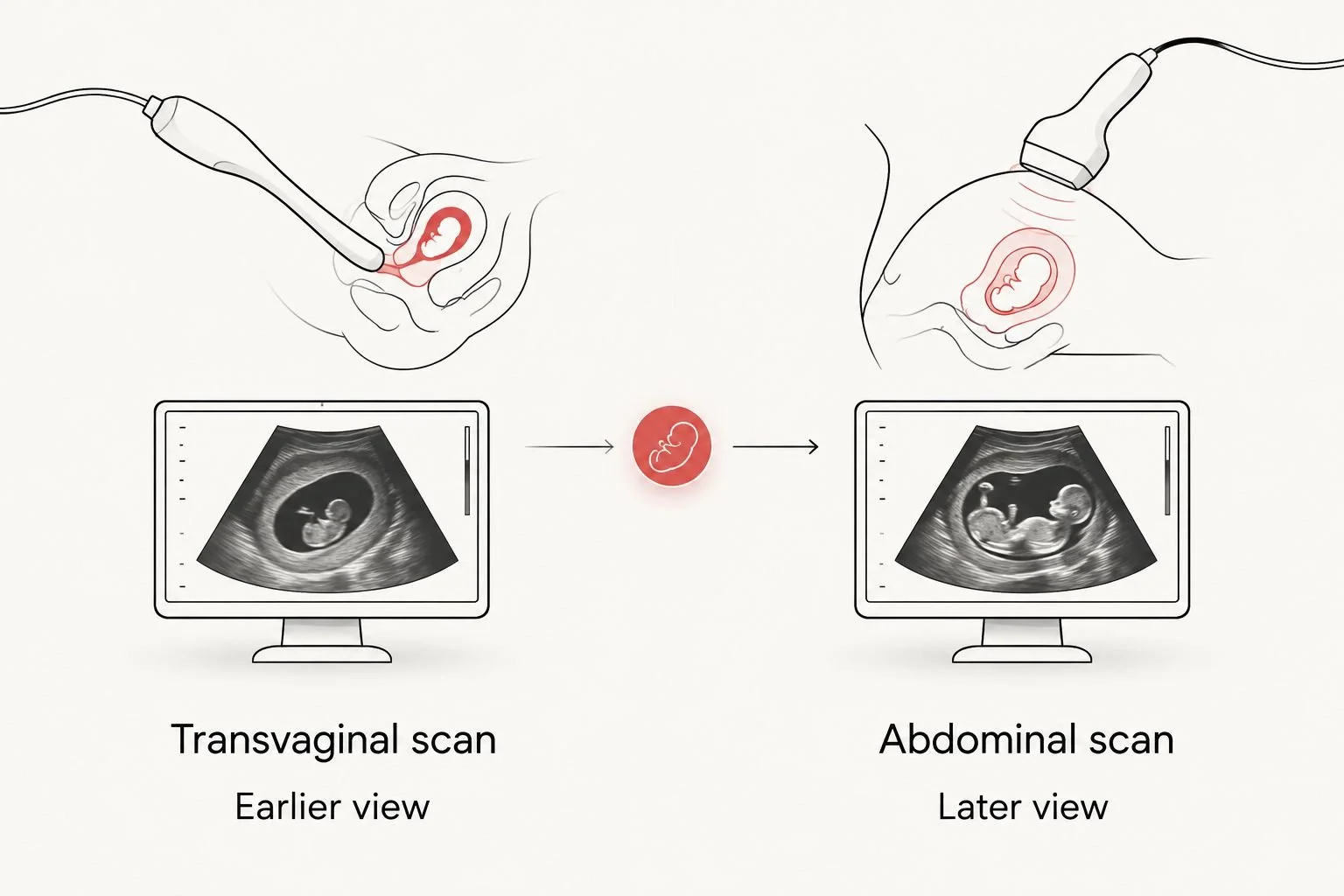
The scan approach affects how early cardiac activity can be identified.
Why the timing varies
There is no single week at which every pregnancy must show visible cardiac activity. When a result differs from what you expected, these are the factors to review with your clinician:
Dating from your last menstrual period. Pregnancy weeks start from the first day of that period. The estimate assumes it reflects when the pregnancy began developing, which is not always precise.
Ovulation timing. Later ovulation can make the pregnancy earlier on ultrasound than the period-based estimate suggests.
How the pregnancy was conceived and dated. If you have fertility-treatment dates, give them to the clinician interpreting the scan rather than relying only on a period estimate.
Scan approach. Transvaginal imaging generally provides more early detail than abdominal imaging.
What the scan actually shows. A sac without a visible embryo calls for a different interpretation from an embryo that has been measured.
The embryo’s measurement. Clinicians use measured findings, not a reported week alone, when assessing whether absent cardiac activity is concerning.
Do not turn these factors into a home checklist for deciding whether a pregnancy is viable. They explain why two people who both say they are 6 weeks pregnant can receive different ultrasound reports. They also explain why the written measurements and the clinician’s interpretation matter more than an isolated image or a brief description from the appointment.
What if the scan shows no heartbeat?
At about 6 weeks, no visible cardiac activity can mean the scan was too early; it can also require clinical follow-up. The finding alone cannot tell you which explanation applies. Ask what was visible, whether an embryo was measured, how the pregnancy was dated and when your clinician recommends reassessment.
There is an important difference between an early scan that cannot establish viability and a scan that meets established diagnostic criteria for pregnancy loss. For example, an embryo with a crown-rump length of 7 millimeters or more and no cardiac activity meets a recognized ultrasound criterion for pregnancy loss when the measurement and examination are reliable. That criterion is based on the embryo’s measured size, not a guess that the pregnancy has reached a particular week. Do not apply it to an image or measurement without a qualified clinician’s interpretation.
A clinician can recommend a repeat ultrasound when the first examination is inconclusive. The interval depends on what the initial scan showed; scheduling another scan immediately is not always the most useful way to answer the question. The follow-up looks for development that could not be confirmed on the first examination.
If you are arranging a reassurance ultrasound between prenatal visits, keep your prenatal clinician involved in interpreting an uncertain result. A private scan can provide images, but an unexpected finding still needs an appropriate clinical follow-up plan.
When to seek care rather than wait for another scan
Do not wait for a routine ultrasound appointment if you have severe or worsening abdominal pain, pain concentrated on one side, shoulder pain, fainting, or heavy bleeding. Seek prompt medical assessment. These symptoms need attention even if an earlier test or scan seemed reassuring.
An early ultrasound question is sometimes also a question about pregnancy location. Before a clinician has confirmed a pregnancy in the uterus, symptoms such as significant one-sided pain or fainting need urgent evaluation for a possible ectopic pregnancy. A keepsake or elective appointment is not a substitute for that assessment.
If you have bleeding or pain that is not severe, contact your prenatal clinician for advice on what to do next. The decision depends on your symptoms and the findings already available. In 2026, the safest use of an early scan is to pair its images with a clear route to clinical interpretation, especially when the answer is uncertain.
How to prepare for an early ultrasound question
Before your appointment, write down the first day of your last menstrual period and any known ovulation or fertility-treatment dates. Bring details of prior scans and reports if you have them. Tell the person arranging the scan whether your goal is pregnancy dating, assessment of symptoms, or an elective look at the pregnancy; those are not interchangeable requests.
At the visit, ask these questions in plain language:
Which scan approach was used? A transvaginal and an abdominal view do not have the same early limits.
What structures were visible? Ask whether the report identifies a gestational sac, an embryo and cardiac activity.
Was the embryo measured? A measured finding carries more clinical meaning than a week estimate alone.
Who will interpret the result? Make sure you know how the report reaches the clinician managing your pregnancy.
What happens if the result is inconclusive? Leave with a follow-up recommendation rather than an unsupported conclusion.
The Ultrasound Concierge is best for patients around Keller and Willow Park who want a private pregnancy ultrasound experience; it is not a replacement for urgent medical evaluation or your prenatal clinician’s care. The Ultrasound Concierge offers both diagnostic and elective ultrasound services, so be clear about which type of assessment you need before the appointment.
Discuss your ultrasound options
Check which pregnancy ultrasound service fits your question and timing.
Can you see a heartbeat at 5 weeks?
At 5 weeks, a heartbeat is generally too early to expect on ultrasound. A scan can show earlier pregnancy structures without yet showing an embryo or cardiac activity. Your clinician should interpret what is visible rather than judge the scan by the week estimate alone.
Is 6 weeks too early for an abdominal ultrasound?
At 6 weeks, an abdominal scan can be too early to show cardiac activity clearly. A transvaginal scan usually offers a more informative early view. An inconclusive abdominal result does not establish that cardiac activity is absent.
When should I repeat an inconclusive early scan?
There is no single repeat-scan date for every inconclusive result. Your clinician sets the timing based on the first scan’s findings, measurements and your clinical situation. Ask for a specific follow-up plan before leaving the appointment.
FAQ
How soon can you see a heartbeat on ultrasound?
Cardiac activity can often be seen at about 6 weeks with a transvaginal scan and around 7 to 8 weeks with an abdominal scan. Pregnancy dating and the structures visible on the scan affect the interpretation.
Can an ultrasound show a heartbeat at 5 weeks?
A heartbeat is generally too early to expect at 5 weeks. Your clinician can explain what pregnancy structures are visible and whether follow-up is needed.
Does no heartbeat at 6 weeks mean miscarriage?
No, absent visible cardiac activity at about 6 weeks does not by itself diagnose pregnancy loss. The scan approach, embryo measurement and dating information matter.
Is a transvaginal scan better than an abdominal scan for an early heartbeat?
A transvaginal scan usually provides a clearer view in the earliest weeks. An abdominal scan often becomes more informative as the pregnancy develops.
Can you see cardiac activity without hearing a heartbeat?
Yes, cardiac activity can appear as visible motion on ultrasound without an audio heartbeat. Hearing sound is not required to identify that motion on the image.
What should I ask after an inconclusive early ultrasound?
Ask which structures were seen, whether an embryo was measured and when your clinician recommends follow-up. Do not interpret an early image without the report and clinical context.
When should I seek urgent care in early pregnancy?
Seek prompt medical assessment for severe or worsening abdominal pain, one-sided pain, shoulder pain, fainting or heavy bleeding. Do not wait for a routine private ultrasound appointment to assess those symptoms.
One last thing
The most useful question after an early scan is often not Was there a heartbeat? but What could this particular scan reliably show? In 2026, an estimate of 6 weeks still cannot replace the scan approach, the measurements and a clinician’s interpretation. The Ultrasound Concierge can be an option for a private pregnancy ultrasound in the Keller and Willow Park area, while your prenatal clinician remains the point of contact for uncertain findings and care decisions.