
Yes, an abdominal ultrasound can detect gallstones and is usually the first imaging test used when a clinician suspects them. It can also show signs of gallbladder inflammation or widened bile ducts, but it does not reliably find every stone, especially stones within the common bile duct. A normal ultrasound does not rule out every cause of gallbladder-related pain.
TL;DR
Can an ultrasound detect gallstones? Yes—abdominal ultrasound is usually the first imaging test for suspected gallbladder stones.
A normal gallbladder ultrasound does not exclude every bile duct stone or gallbladder function problem.
Severe abdominal pain with fever, jaundice, or persistent vomiting needs urgent medical assessment.
The Ultrasound Concierge suits Keller and Willow Park patients seeking private diagnostic ultrasound; confirm the specific examination before booking.
Why this matters: Finding stones and explaining your pain are different tasks. Your clinician considers the scan alongside your symptoms, examination, and sometimes blood tests; an elective imaging visit does not replace that assessment. Before arranging diagnostic imaging in 2026, review whether you need a doctor’s referral for a private ultrasound and confirm the requirements for your examination.
Can an ultrasound detect gallstones?
Yes. Ultrasound is particularly useful for finding stones inside the gallbladder. The National Institute of Diabetes and Digestive and Kidney Diseases identifies ultrasound as the best imaging test for finding gallstones. The examination uses sound waves rather than ionizing radiation.
A sonographer places gel on your upper abdomen and moves a handheld probe over the area. Gallstones commonly appear as bright structures that cast a dark shadow behind them, and many move when you change position. The examiner also checks the surrounding structures rather than looking for stones alone.
Your report can describe:
Stones: Their appearance and location within the gallbladder.
Sludge: Thickened bile containing material that can collect inside the gallbladder.
Gallbladder wall changes: Thickening that needs interpretation alongside other findings.
Surrounding fluid: A possible sign of inflammation in the appropriate clinical setting.
Bile duct size: Widening that can raise concern about impaired bile drainage.
None of these findings should be interpreted in isolation. For example, gallbladder wall thickening has causes other than gallbladder inflammation, while a stone found incidentally does not automatically explain abdominal discomfort.
What can an ultrasound miss?
Ultrasound has limits even when the examination is performed correctly. Very small stones can be difficult to see, and bowel gas can obscure parts of the biliary system. A report describing limited visualization means the examiner could not fully assess a structure—not that the structure is definitely normal.
Stones in the common bile duct
The common bile duct carries bile toward the intestine. A stone that leaves the gallbladder can become lodged there, but an abdominal ultrasound does not consistently show the entire duct clearly.
Sometimes the scan reveals an indirect clue, such as duct widening, without showing the stone itself. Your clinician can use symptoms, liver blood tests, and additional imaging to decide whether a blocked duct needs further investigation.
Problems with gallbladder emptying
A standard ultrasound shows anatomy; it does not directly measure how well the gallbladder empties. You can have symptoms that warrant further assessment even when no stones are visible.
A clinician sometimes requests a hepatobiliary scan, commonly called a HIDA scan, to answer a functional question. A normal ultrasound alone does not establish or exclude a functional gallbladder disorder.
Other causes of upper abdominal pain
Pain beneath the right ribs is not unique to gallstones. Problems involving the stomach, liver, pancreas, kidneys, lungs, or muscles can produce overlapping symptoms.
Persistent symptoms deserve reassessment, not repeated assumptions that a normal scan settles the diagnosis. Tell your clinician whether the pain changes, spreads, or occurs with fever, vomiting, or yellowing of the skin or eyes.
Which test is best for suspected gallstones?
For a patient evaluated in 2026, the useful distinction is what question each test answers—not which examination sounds most advanced. Abdominal ultrasound usually comes first for suspected gallbladder stones. Other tests address unresolved questions about the bile ducts or gallbladder function.
Abdominal ultrasound
Best for: Initial evaluation of suspected gallbladder stones
Main advantage: Shows gallstones and surrounding structures without ionizing radiation
Main limitation: Bowel gas and limited views can hide abnormalities, especially in the bile duct
MRCP
Best for: Detailed assessment of the bile ducts when obstruction remains a concern
Main advantage: Uses MRI to produce detailed images of the biliary system
Main limitation: Does not remove a stone or provide treatment
HIDA scan
Best for: Selected questions about bile flow, acute gallbladder inflammation, or gallbladder function
Main advantage: Assesses movement of a radiotracer through the biliary system
Main limitation: Uses a radioactive tracer and is not the usual first test for finding gallstones
These tests are not interchangeable. MRCP stands for magnetic resonance cholangiopancreatography; despite the long name, its purpose is straightforward: to examine the bile and pancreatic ducts in detail.
A HIDA scan answers a different type of question. Your clinician chooses it based on the suspected condition and the findings already available, rather than simply because an ultrasound was negative.
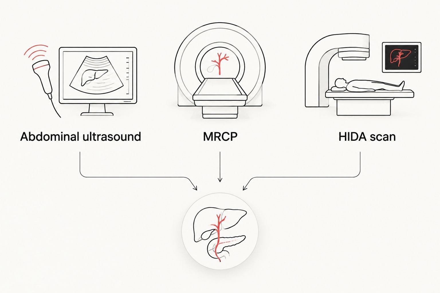
The right examination depends on whether the question concerns stones, bile ducts, or gallbladder function.
Why gallstone detection varies
A clear ultrasound depends on both the examination conditions and the location of the suspected problem. These factors explain why one patient receives a definite answer while another needs further assessment:
Stone location: Stones inside the gallbladder are generally easier to identify than stones hidden within the common bile duct.
Stone size and appearance: Tiny stones and thickened bile can be harder to distinguish than a clearly shadowing stone.
Food before the scan: Eating causes the gallbladder to contract, which can make it harder to examine.
Bowel gas: Gas interferes with ultrasound transmission and can block the view of deeper structures.
Body tissues and anatomy: The depth and position of the gallbladder affect how clearly sound waves depict it.
Examination scope: A targeted right-upper-abdominal examination and a scan ordered for another body area do not answer the same question.
You do not need to judge image quality yourself. Ask whether the gallbladder and bile ducts were adequately visualized and whether the report identifies any limitations that change the next step.
How should I prepare for a gallbladder ultrasound?
Follow the imaging provider’s instructions for your specific examination. RadiologyInfo’s abdominal ultrasound guidance describes avoiding food for 8–12 hours before some examinations of the liver, gallbladder, spleen, and pancreas. Your own instructions take priority over a general preparation window.
Do not stop prescribed medicines, change diabetes treatment, or restrict fluids without checking with the appropriate clinician. If fasting is difficult or medically complicated, contact the imaging provider before the appointment rather than improvising.
Use this preparation sequence for a diagnostic appointment in 2026:
Confirm the examination. Ask whether the request is for a gallbladder, right-upper-quadrant, or complete abdominal ultrasound.
Confirm preparation. Ask exactly when to stop eating and what the instructions say about water and medicines.
Share your history. Mention previous gallstones, gallbladder surgery, relevant symptoms, and earlier imaging.
Confirm follow-up. Know which clinician receives the report and who will explain the findings to you.
If you have already eaten, tell the imaging team. They can decide whether the examination should proceed or be rescheduled; you do not need to make that decision alone.
What happens during the examination?
You usually lie on an examination table while the sonographer scans your upper abdomen. You might be asked to turn onto your side, take a deep breath, or briefly hold your breath so the gallbladder can be viewed from different angles.
RadiologyInfo states that an abdominal ultrasound is usually completed within 30 minutes. The actual examination depends on the requested study and how easily the structures can be seen; completing the images is separate from receiving the interpreted report.
The probe can cause discomfort when it presses over an already tender area. Tell the sonographer where it hurts and whether the pressure becomes difficult to tolerate. Tenderness during scanning is information for the clinical assessment, not a diagnosis by itself.
Does finding gallstones mean I need surgery?
Finding gallstones does not automatically mean you need surgery. NIDDK explains that gallstones without symptoms usually do not need treatment. Your clinician considers whether the stones are causing symptoms or complications before recommending management.
If you have recurrent attacks or evidence of inflammation or obstruction, the next step differs from an incidental finding on an unrelated scan. Do not use the number or apparent size of stones alone to decide how serious the situation is.
For your 2026 follow-up visit, bring a short description of the pain: where it starts, how long it lasts, whether it follows eating, and what other symptoms occur. This history helps your clinician judge whether the imaging findings fit your experience.
When should I seek urgent care instead of booking a scan?
Seek urgent medical assessment for severe or persistent abdominal pain, particularly with fever, chills, jaundice, or repeated vomiting. Gallstones can cause inflammation, infection, or blocked bile drainage, and a routine outpatient appointment is not a substitute for evaluating those symptoms.
Dark urine or pale stools alongside abdominal pain also need prompt medical attention. If you feel seriously unwell or symptoms are rapidly worsening, use emergency care rather than waiting for an elective imaging visit.
Do not delay urgent assessment to finish a fasting period. Tell the treating team when you last ate or drank and let them direct the evaluation.
Choosing a private diagnostic ultrasound appointment
The Ultrasound Concierge is best for Keller and Willow Park patients seeking private diagnostic ultrasound in a personalized setting. The practice provides diagnostic and elective imaging, but those categories serve different purposes. A pregnancy keepsake scan does not replace a diagnostic examination for abdominal symptoms.
Before contacting The Ultrasound Concierge, clarify the examination your clinician wants. Confirm whether the practice performs that specific study, what documentation it requires, how to prepare, and how the report reaches your treating clinician.
A calm setting supports the visit, but it does not remove ultrasound’s limitations or replace medical follow-up. If your symptoms suggest an urgent problem, seek clinical assessment first.
FAQ
Can an ultrasound detect gallstones even if I am not having pain?
Yes, an ultrasound can detect gallstones when you are not having pain. Stones are sometimes found incidentally, and finding them does not automatically mean treatment is needed.
Can an ultrasound miss a gallstone?
Yes, an ultrasound can miss a gallstone, particularly a very small stone or one within the common bile duct. Persistent symptoms or abnormal blood tests can justify further assessment despite a negative scan.
Do I need to fast before a gallbladder ultrasound?
Fasting is commonly requested before a gallbladder ultrasound. RadiologyInfo describes an 8–12-hour period without food for some upper abdominal examinations, but follow your imaging provider’s instructions about food, fluids, and medicines.
Is an ultrasound painful when you have gallstones?
An ultrasound can be uncomfortable when the probe presses over a tender area. Tell the sonographer about pain so the examination can be adjusted appropriately.
Can a gallbladder ultrasound show whether a stone is blocking a duct?
A gallbladder ultrasound can show signs of a blocked bile duct, such as duct widening, but it does not reliably show every duct stone. Your clinician can request further testing when obstruction remains a concern.
Does a normal ultrasound mean my gallbladder is healthy?
A normal ultrasound does not exclude every gallbladder or bile duct problem. It does not directly measure gallbladder emptying, and limited views can leave questions unresolved.
Can The Ultrasound Concierge help me arrange diagnostic imaging?
The Ultrasound Concierge provides private diagnostic ultrasound services for patients in the Keller and Willow Park area. Confirm the specific abdominal examination, preparation requirements, and reporting process before arranging a visit.
One last thing
Ask whether the scan explains your symptoms—not just whether it found stones. An incidental gallstone and a normal but limited examination can both require a more thoughtful conversation than a simple positive or negative result. For any ultrasound arranged in 2026, leave with a clear plan for who reviews the report and what happens if the pain continues.
Medical background: NIDDK’s patient guidance on gallstone diagnosis and treatment, RadiologyInfo’s abdominal ultrasound guidance, and the American College of Radiology’s Appropriateness Criteria for right upper quadrant pain inform the general explanations above. This article provides education, not an individual diagnosis.