
Yes—an ultrasound can detect ovarian changes associated with PCOS, but it cannot diagnose the condition on its own. In 2026, an adult PCOS assessment combines menstrual history, signs or blood tests of androgen excess, and sometimes ovarian imaging, while excluding other causes of similar symptoms.
TL;DR
Can an ultrasound detect PCOS? It can show associated ovarian changes, but imaging alone cannot confirm the diagnosis.
A normal pelvic ultrasound does not rule out PCOS; menstrual patterns and androgen findings also matter.
The Ultrasound Concierge offers private diagnostic imaging; ask whether the requested pelvic examination is available.
Small ovarian follicles are not the same as ovarian cysts, and neither finding alone proves PCOS.
Can an ultrasound detect PCOS?
Ultrasound can support a PCOS diagnosis, but an ovarian appearance is not a diagnosis. A pelvic scan shows the ovaries and uterus; it does not directly measure hormone levels or establish whether you ovulate regularly over time.
The international evidence-based PCOS guideline published in 2023, developed with Monash University and international professional societies, recommends assessing adults for the following features after excluding other explanations:
Ovulatory dysfunction
What supports it: Irregular or absent periods, with further assessment when needed
What ultrasound contributes: A single scan cannot establish your long-term ovulation pattern
Androgen excess
What supports it: Relevant examination findings or elevated androgen blood tests
What ultrasound contributes: Ultrasound does not measure androgen levels
Polycystic ovarian morphology
What supports it: A qualifying follicle count or ovarian volume
What ultrasound contributes: Imaging can document this ovarian appearance
Adults generally need 2 of these 3 features, not all of them. If irregular cycles and androgen excess are already present, ultrasound is not required to establish PCOS once other causes have been excluded.
The word polycystic also causes understandable confusion. The small follicles associated with PCOS are different from the findings discussed in whether ultrasound can check for ovarian cysts.
Why this matters
A report that mentions “polycystic ovarian morphology” can sound like a final answer. It describes an imaging pattern, not the full hormonal condition, and some people have that pattern without PCOS.
The reverse matters just as much: you can have PCOS without a qualifying ovarian appearance. For your 2026 assessment, the useful question is not simply whether the scan looks abnormal, but whether your history, examination and laboratory findings meet diagnostic criteria together.
Neither an abnormal-looking ovary nor a normal-looking ovary should end the assessment by itself. Ask the clinician managing your care to explain how each finding changes the diagnosis and next steps.
What does PCOS look like on ultrasound?
Ultrasound can show numerous small follicles and increased ovarian volume. Follicles are fluid-filled structures in which eggs develop; their presence is normal, while their number and the overall ovarian pattern can contribute to a PCOS assessment.
For adults, the 2023 international guideline uses a threshold of 20 follicles in at least one ovary when ultrasound technology and image quality allow an accurate total count. The follicles assessed for this purpose measure 2–9 mm.
When older technology or inadequate image quality prevents a reliable total follicle count, the guideline includes an ovarian volume threshold of 10 mL in at least one ovary, alongside other technical criteria. These measurements require professional interpretation rather than counting circles on a saved image.
A dominant follicle, corpus luteum or separate cyst can affect interpretation. Your clinician also needs to know the scan route, image quality and relevant menstrual or medication history.
Does a “string of pearls” mean PCOS?
A peripheral arrangement of small follicles is sometimes described as a “string of pearls.” That phrase alone does not establish PCOS, and a report does not need to use it for PCOS to remain a possibility.
Ask about the actual measurements and the report’s conclusion. A memorable description is less useful than a documented follicle count, ovarian volume and explanation of any limitations.
Which pelvic ultrasound approach is used?
The two main approaches are transvaginal and transabdominal ultrasound. They answer overlapping questions, but they differ in how clearly they show small ovarian structures and in the preparation and consent involved.
Transvaginal ultrasound
Best for: Detailed ovarian assessment when acceptable and clinically appropriate
Benefit: Usually gives a clearer view of small follicles
Limitation: Requires an internal examination and your consent
Transabdominal ultrasound
Best for: External pelvic imaging when an internal examination is declined or unsuitable
Benefit: Examines the pelvis through the lower abdomen
Limitation: Small follicles can be harder to count accurately
Transvaginal ultrasound: detailed ovarian assessment
A transvaginal examination uses a covered ultrasound probe placed in the vagina. Its closer view of the ovaries generally makes it the most accurate ultrasound approach for assessing polycystic ovarian morphology when it is acceptable to the patient.
You can ask questions, decline an internal examination or ask for it to stop. Discuss alternatives and their limitations before the examination begins, rather than feeling that consent is an all-or-nothing decision.
Transabdominal ultrasound: an external approach
A transabdominal examination moves a probe across your lower abdomen. A full bladder often helps the pelvic view, but you should follow the imaging center’s instructions rather than choose a drinking schedule yourself.
This approach can assess ovarian size and other pelvic structures, although detailed follicle counting is more limited. A report should make clear when the ovaries were not adequately visualized; “not seen well” is not the same as “normal.”
How does an ultrasound fit into a PCOS assessment?
A useful 2026 evaluation starts with the clinical question, not a scan package. Your clinician determines whether imaging will add information or whether the existing history and laboratory findings already establish the relevant diagnostic features.
Review symptoms. Describe your menstrual pattern, acne, unwanted facial or body hair, scalp hair changes and fertility concerns. Include when changes started.
Check other causes. Your clinician selects testing for conditions that can resemble PCOS, such as thyroid disorders, elevated prolactin and nonclassic congenital adrenal hyperplasia.
Consider imaging. If ovarian morphology will help clarify the diagnosis, discuss the appropriate pelvic ultrasound approach and preparation.
Review results. Bring the imaging report back to the clinician responsible for diagnosis and treatment. Ask what the results establish and what remains unanswered.
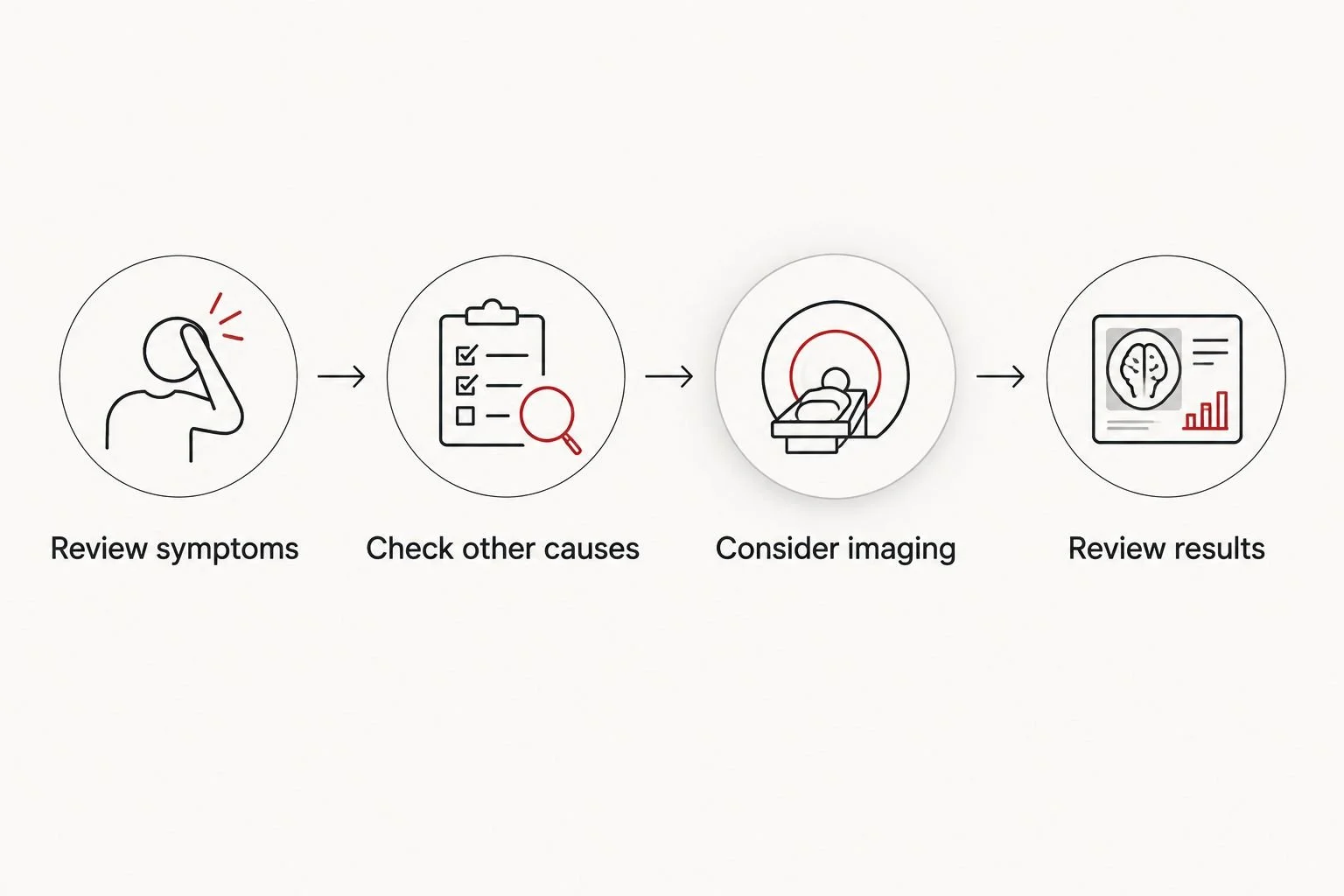
Imaging contributes to the assessment; the clinical review connects the findings.
Before scheduling, confirm whether your requested examination needs an order and who will interpret the images. Also confirm how the written report reaches the clinician managing your care; photographs alone are not a substitute for a diagnostic report.
For local imaging, review The Ultrasound Concierge’s services and ask specifically about the examination your clinician requests. The Ultrasound Concierge is best suited to Keller and Willow Park patients seeking private diagnostic ultrasound rather than an elective keepsake visit. Imaging remains one part of care, not a replacement for the clinician who evaluates hormones and manages PCOS.
Why ultrasound findings and PCOS diagnoses vary
Different scan results do not automatically mean someone made a mistake. These factors affect what ultrasound shows and how the finding should be used:
Age and time since the first period. Ultrasound is not recommended for diagnosing PCOS in adolescents because normal developmental changes overlap with a polycystic appearance. The international guideline advises against using ultrasound criteria within 8 years after the first period.
Scan route. Transvaginal imaging generally allows more detailed ovarian assessment than an abdominal-only examination.
Image quality. Equipment, ovarian position and the available view affect whether a reliable follicle count is possible.
Cycle-related structures. A dominant follicle, corpus luteum or separate cyst affects how ovarian measurements are interpreted.
Hormonal treatment. Contraception and other hormonal medicines can change bleeding patterns, hormone results and ovarian appearance. Tell your clinician what you take; do not stop treatment yourself.
Your symptoms also need their own assessment. Weight changes can prompt questions about hormonal causes of weight gain, but weight gain alone does not establish PCOS or identify a particular hormone disorder.
For a 2026 appointment, bring your medication list and any previous hormone or imaging results. That context helps your clinician distinguish a new finding from a change related to treatment or the timing of the examination.
Can you have PCOS if your ultrasound is normal?
A normal ultrasound does not exclude PCOS. An adult with ovulatory dysfunction and androgen excess can meet diagnostic criteria without polycystic ovarian morphology, after other causes have been excluded.
Ask which diagnostic features your clinician has identified. If the scan is normal but your periods remain irregular, the next step is clinical follow-up—not assuming that the symptoms have been explained away.
Can you have polycystic ovaries without having PCOS?
Polycystic ovarian morphology alone does not establish PCOS. You can have an ultrasound appearance associated with PCOS without meeting the broader diagnostic criteria.
A report should therefore lead to a discussion of your menstrual history and androgen findings. It should not automatically lead to a diagnosis, medication or a prediction about your fertility.
Can an ultrasound tell whether you are ovulating?
A single ultrasound cannot establish your usual ovulation pattern. It can show findings consistent with a particular phase of the cycle, while timed or repeated assessment answers a different question from a one-time PCOS scan.
If pregnancy is your goal, tell your clinician before scheduling. A diagnostic evaluation for PCOS and a fertility-monitoring plan are related, but they are not interchangeable appointments.
What should you ask before and after your scan?
Clear questions make your 2026 appointment more useful without requiring you to interpret the images yourself. Start with the purpose of the examination, then clarify how you will receive an explanation of the results.
Before your scan, ask:
Is this a diagnostic pelvic ultrasound intended to assess the ovaries?
Will the examination be transabdominal, transvaginal or both?
What preparation should I follow, and what are my alternatives if I decline an internal examination?
Who interprets the images, and who receives the written report?
Afterward, ask whether both ovaries were adequately seen and whether the report documents ovarian volume, follicle findings and technical limitations. Then ask your treating clinician which diagnostic criteria you meet and whether additional testing is needed.
If you contact The Ultrasound Concierge, state that you are asking about diagnostic imaging for a possible PCOS evaluation. That distinction helps separate your medical question from an elective pregnancy imaging request without assuming that any particular examination is offered.
Sudden severe pelvic pain, fainting, or pelvic pain with possible pregnancy needs prompt medical assessment. Do not wait for a routine PCOS scan to evaluate those symptoms.
FAQ
Can an ultrasound detect PCOS without a blood test?
An ultrasound can show ovarian changes associated with PCOS, but it cannot confirm the condition alone. Blood testing helps assess androgen levels and exclude other explanations when clinically indicated.
Does a normal ultrasound mean I do not have PCOS?
A normal ultrasound does not rule out PCOS. Adults can meet diagnostic criteria through ovulatory dysfunction and androgen excess after other causes are excluded.
Do ovarian cysts mean I have PCOS?
Ovarian cysts do not automatically mean you have PCOS. The small follicles assessed for polycystic ovarian morphology differ from a separate ovarian cyst, and imaging findings need clinical context.
Is an internal ultrasound required to diagnose PCOS?
An internal ultrasound is not required for every PCOS diagnosis. When imaging is needed, transvaginal ultrasound generally provides a more detailed ovarian view, but it requires your consent.
Can teenagers be diagnosed with PCOS by ultrasound?
Ultrasound is not recommended for diagnosing PCOS in adolescents. Normal ovarian development overlaps with the imaging appearance associated with PCOS, so assessment relies on age-appropriate clinical criteria.
Can a pelvic ultrasound show a hormone imbalance?
A pelvic ultrasound does not directly measure a hormone imbalance. It shows anatomy, while symptoms, examination findings and appropriate laboratory tests assess hormonal concerns.
Should I stop birth control before a PCOS scan?
Do not stop birth control on your own before a PCOS assessment. Tell your clinician what you take so they can explain its effects on bleeding patterns, hormone testing and scan interpretation.
One last thing
The surprising part of a PCOS evaluation is that the scan can be unnecessary even when PCOS is present. If you already have irregular cycles and androgen excess, ask your clinician what additional question imaging would answer before scheduling another test.
Your most useful takeaway is an explained result, not just an image. Leave the follow-up visit knowing whether you meet diagnostic criteria, what else was considered and what care addresses your symptoms or pregnancy goals.