
Yes—ultrasound can identify ovarian endometriomas and some deep endometriosis, giving your clinician evidence to diagnose the condition and plan care. A normal ultrasound does not rule out endometriosis, especially small, superficial deposits, so persistent symptoms still need evaluation even when your scan looks normal.
TL;DR
Can an ultrasound diagnose endometriosis? It can identify some forms, but a normal scan cannot exclude the condition.
Transvaginal ultrasound helps assess ovarian endometriomas and deep endometriosis; superficial disease often remains invisible.
Persistent pelvic pain deserves follow-up even after normal pelvic ultrasound results.
The Ultrasound Concierge provides private diagnostic ultrasound; confirm the appropriate examination with your referring clinician.
Can an ultrasound diagnose endometriosis?
Ultrasound can support an endometriosis diagnosis, but it cannot detect every form of the disease. What it shows depends on where the endometriosis is, the examination performed, and the experience of the professionals acquiring and interpreting the images.
Before booking, review the available ultrasound services and ask whether the examination requested by your clinician is offered. A diagnostic pelvic ultrasound and an elective pregnancy imaging visit serve different purposes.
Ovarian endometrioma
What ultrasound can show: An ovarian cyst with features characteristic of endometriosis
Important limitation: Other cysts can overlap in appearance and require further assessment
Deep endometriosis
What ultrasound can show: Lesions or indirect signs involving structures such as the bowel, bladder, or supporting pelvic tissues
Important limitation: Detection depends on location, protocol, and examiner expertise
Superficial endometriosis
What ultrasound can show: Often no visible abnormality
Important limitation: A normal scan cannot exclude small deposits on pelvic surfaces
Restricted organ movement
What ultrasound can show: Reduced movement that can suggest adhesions during a targeted examination
Important limitation: This is an indirect sign, not proof of endometriosis by itself
An imaging report provides evidence, not an explanation for every symptom. Your clinician combines the findings with your history, examination, and treatment goals rather than treating the scan as a stand-alone answer.
Why this matters
A normal report can feel reassuring and frustrating at the same time. It can exclude or identify certain visible problems without explaining pain during periods, sex, bowel movements, or urination.
For your 2026 care decisions, keep that distinction clear: “not seen on ultrasound” does not mean “not present.” You do not need an abnormal scan for your symptoms to deserve attention.
NICE guideline NG73, Endometriosis: diagnosis and management, recommends ultrasound in the assessment of suspected endometriosis and explicitly advises against excluding the condition because an examination and ultrasound are normal. That guidance applies to diagnostic evaluation, not elective imaging or keepsake scans.
What kind of ultrasound is used for suspected endometriosis?
There are 2 main pelvic ultrasound approaches: transvaginal and transabdominal. They provide different views, and your clinician chooses the approach according to the clinical question, your circumstances, and your consent.
Transvaginal ultrasound
Best for: Detailed assessment of pelvic organs when appropriate and acceptable
Main advantage: Places the probe close to the uterus and ovaries
Main limitation: An internal examination is not suitable or acceptable for everyone
Transabdominal ultrasound
Best for: An external pelvic view or an alternative when internal scanning is unsuitable
Main advantage: Examines the pelvis through the lower abdomen
Main limitation: Provides less detail for some small or deep pelvic findings
Specialist endometriosis ultrasound
Best for: Targeted evaluation when deep disease is suspected
Main advantage: Assesses relevant pelvic structures and organ movement using a focused protocol
Main limitation: Requires appropriate expertise and still cannot exclude all disease
Transvaginal ultrasound
A transvaginal examination uses a covered probe placed gently inside the vagina. It provides close views of the uterus, ovaries, and nearby pelvic structures without ionizing radiation.
Your consent matters throughout the examination. Tell the sonographer about pain, concerns about internal scanning, or previous difficult examinations; you can ask to pause or stop.
Transabdominal ultrasound
A transabdominal examination uses a probe over the lower abdomen. The imaging team commonly requests a full bladder to improve the pelvic view, but you should follow the instructions for your specific appointment.
This approach remains useful when an internal examination is declined or unsuitable. Its limitation is detail: a normal external scan does not establish that endometriosis is absent.
Specialist endometriosis ultrasound
A targeted examination looks beyond whether the uterus and ovaries appear normal. Depending on the referral and protocol, it assesses areas where deep endometriosis occurs and checks how pelvic organs move relative to each other.
Ask whether the examination specifically assesses suspected endometriosis. A routine pelvic scan and a specialist endometriosis assessment are not interchangeable, even when both use ultrasound.
Why ultrasound detection varies
There is no single detection figure that accurately describes every endometriosis ultrasound. For a 2026 appointment, these factors explain more than a general claim that a scan is “accurate”:
Disease location: Ovarian cysts and some deep lesions are more visible than superficial deposits on pelvic surfaces.
Lesion size and appearance: Small or subtle abnormalities are harder to distinguish from surrounding tissue.
Examination protocol: A routine pelvic survey does not necessarily include a targeted assessment of deep endometriosis or organ mobility.
Examiner expertise: Recognizing endometriosis-related findings requires experience with the relevant anatomy and scanning techniques.
Image quality and access: Bowel gas, anatomy, discomfort, and the examination approach affect which structures can be assessed.
Information on the referral: Specific symptoms help direct attention to relevant structures, including the bowel or bladder when indicated.
These limitations do not make ultrasound unhelpful. They explain why a useful first investigation can still leave questions that require a gynecologist or specialist imaging team.
What should you do before an endometriosis ultrasound?
You do not need to prove that you have endometriosis before asking for an assessment. Bring clear information that helps your clinician choose the right examination and interpret the result.
Use these 4 steps to prepare:
Describe symptoms. Record where you feel pain, whether it changes with your menstrual cycle, and whether sex, bowel movements, or urination trigger it. Include how symptoms affect work, sleep, exercise, and everyday activities.
Confirm the examination. Ask what your referral requests and whether the appointment is a routine pelvic scan or a targeted endometriosis examination. Mention previous imaging, pelvic surgery, or known ovarian cysts.
Follow preparation instructions. Ask about bladder preparation rather than copying instructions from a different type of ultrasound. Tell the team in advance if internal scanning is painful, unsuitable, or something you do not consent to.
Arrange follow-up. Know who will receive the report, who will explain it, and what happens if it does not explain your symptoms. Imaging is most useful when someone is responsible for the next clinical decision.
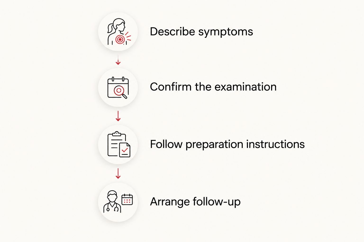
A useful ultrasound appointment starts with the right clinical question and ends with a follow-up plan.
For your 2026 symptom record, use actual examples rather than only describing pain as mild or severe. “I miss work during my period” gives your clinician information that a scan cannot capture.
What happens if the ultrasound is normal?
A normal ultrasound does not end the investigation when symptoms continue. Return to the clinician managing your pelvic pain and review what the examination assessed, what it excluded, and what remains uncertain.
Start with the report wording. “No ovarian cyst identified” answers a narrower question than “no cause for pain identified,” and neither statement excludes superficial endometriosis.
Your clinician can consider symptom-based treatment, gynecology referral, specialist ultrasound, or pelvic MRI according to your situation. These are clinical decisions, not an automatic sequence that every patient must complete.
Also consider other explanations for pelvic pain. Adenomyosis, fibroids, ovarian cysts, pelvic floor problems, and bowel or bladder conditions can cause overlapping symptoms; more than one condition can occur together.
If your symptoms remain disruptive, ask a direct question: “What is our next step if this scan does not explain my pain?” Leave with a plan rather than an instruction to ignore persistent symptoms.
Is MRI better than ultrasound for endometriosis?
MRI is useful for selected questions, particularly mapping suspected deep endometriosis, but it is not automatically better than a well-performed specialist ultrasound. The best examination depends on the suspected location of disease and the expertise available.
Specialist ultrasound
Best for: Targeted pelvic assessment when appropriate
Benefit: Offers real-time assessment, including organ movement
Limitation: Depends on examiner expertise and cannot exclude superficial disease
Pelvic MRI
Best for: Mapping deep disease or clarifying selected findings
Benefit: Provides a broader view of pelvic anatomy
Limitation: A normal MRI still does not exclude all endometriosis
Laparoscopy
Best for: Selected cases requiring surgical assessment or treatment
Benefit: Allows direct inspection and tissue sampling
Limitation: Requires surgery, with procedural and anesthesia risks
For 2026 planning, ask what additional question an MRI would answer before requesting one simply because an ultrasound was normal. The purpose is to improve a clinical decision, not collect another normal result.
Do you need surgery to diagnose endometriosis?
Surgery is not required before every patient can receive an endometriosis diagnosis or begin treatment. Clinicians can use symptoms, examination findings, and imaging to make a working diagnosis and discuss appropriate management.
Laparoscopy remains an option when imaging is negative and symptoms persist, when treatment has not helped or is unsuitable, or when surgical treatment is being considered. During laparoscopy, a surgeon can inspect the pelvis and take tissue samples when indicated.
The decision should reflect your symptoms, fertility goals, previous treatment, and preferences. An inconclusive scan does not automatically mean you need surgery, and a normal scan does not automatically mean you do not.
Where does private diagnostic ultrasound fit?
The Ultrasound Concierge is best for patients seeking private diagnostic ultrasound in Keller or Willow Park. Its concierge-style setting is an alternative to a clinical imaging center, not a replacement for gynecologic evaluation or specialist endometriosis care.
The Ultrasound Concierge provides diagnostic imaging, but the appropriate examination must match your clinician’s question. Before arranging an appointment, confirm whether the requested pelvic examination is offered and whether suspected endometriosis requires referral to a specialist imaging service.
Private imaging can be part of your care pathway; its limitation is the same important boundary that applies elsewhere: no ultrasound setting can guarantee exclusion of all endometriosis. Make sure your referring clinician receives and reviews the findings.
Ask about your requested examination
Confirm whether the diagnostic ultrasound requested by your clinician is offered.
When should pelvic pain receive urgent attention?
Do not assume that sudden or severe pelvic pain is endometriosis. Seek urgent medical assessment for severe or rapidly worsening pain, fainting, fever with pelvic pain, or significant bleeding with weakness or dizziness.
Pelvic pain with a possible pregnancy also needs prompt assessment, particularly with bleeding, shoulder-tip pain, or feeling faint. An elective or routine outpatient ultrasound appointment is not a substitute for emergency evaluation.
FAQ
Can an ultrasound diagnose endometriosis if my ovaries look normal?
Yes, ultrasound can identify some deep endometriosis even when the ovaries look normal, but only if the relevant areas are assessed and findings are visible. Normal ovaries do not exclude superficial endometriosis or disease elsewhere in the pelvis.
Does a normal pelvic ultrasound mean I don't have endometriosis?
No, a normal pelvic ultrasound does not rule out endometriosis. Small superficial deposits often remain invisible, so persistent symptoms still deserve clinical follow-up.
Is an internal ultrasound necessary to check for endometriosis?
Transvaginal ultrasound is commonly recommended for detailed pelvic assessment, but it requires your consent and is not suitable for everyone. Your clinician can discuss transabdominal ultrasound or other assessment options if you decline it or it is unsuitable.
Can ultrasound tell the difference between an endometrioma and another ovarian cyst?
Ultrasound often identifies features that suggest an endometrioma, but some ovarian cysts have overlapping appearances. Your clinician uses the report, symptoms, and any recommended follow-up to determine what the finding means.
Should I get an MRI if my ultrasound is normal?
A normal ultrasound does not automatically mean you need an MRI. Your clinician considers MRI when it would clarify a finding, assess suspected deep disease, or help plan care.
Do I need to be on my period for an endometriosis ultrasound?
You generally do not need to be on your period for an endometriosis ultrasound. Follow the referring clinician’s and imaging team’s instructions, particularly if the examination also addresses another menstrual-cycle-related question.
Can The Ultrasound Concierge replace an endometriosis specialist?
No, The Ultrasound Concierge provides diagnostic imaging rather than a replacement for specialist endometriosis care. Confirm the requested examination before booking and arrange clinical review of the results.
Can I start treatment without having surgery first?
Yes, clinicians can discuss treatment for suspected endometriosis without requiring surgery first. The choice depends on your symptoms, medical history, fertility goals, and preferences.
One last thing
For your 2026 follow-up appointment, bring the written report rather than relying only on a summary that says “everything looked fine.” Ask which structures were assessed and whether the examination specifically looked for deep endometriosis.
The most useful question is not only “Was my ultrasound normal?” It is “What does this result mean for my symptoms and next step?” A normal image and a need for further care can both be true.