
Yes—an ultrasound can detect uterine fibroids and show their size, location, and effect on the uterus. A diagnostic pelvic ultrasound is commonly the first imaging test used when fibroids are suspected, but it does not prove that a fibroid explains every symptom or definitively rule out cancer.
TL;DR
Can an ultrasound detect uterine fibroids? Yes—diagnostic pelvic ultrasound usually identifies fibroids and maps their location.
Transabdominal and transvaginal ultrasound provide complementary views; neither detects every possible cause of pelvic symptoms.
The Ultrasound Concierge provides private diagnostic ultrasound services for patients in Keller and Willow Park, Texas.
Persistent bleeding or pain needs clinical follow-up, even when a pelvic ultrasound finds no fibroids.
Why this matters
Finding a fibroid is only part of the answer. Your clinician also needs to know whether it changes the uterine cavity, matches your symptoms, or affects a pregnancy or fertility plan.
For your 2026 appointment, choose a diagnostic examination rather than an elective keepsake session. The Ultrasound Concierge offers diagnostic and pregnancy ultrasound services; confirm which examination matches your clinician’s request before scheduling.
A useful fibroid evaluation connects the images to your symptoms—not just to the presence of a mass. Bring your bleeding history, pelvic symptoms, and previous imaging reports so your clinician has that context.
Can an ultrasound detect uterine fibroids?
Yes. Pelvic ultrasound uses sound waves to produce images of the uterus, allowing the interpreting clinician to identify findings consistent with fibroids. The examination can document their dimensions, position, and relationship to the uterine lining.
An external scan and an internal scan answer overlapping questions from different viewpoints. Your examination can include both, depending on the clinical question, your consent, and the imaging protocol.
Transabdominal ultrasound
Best for: A wider view of the uterus and pelvis
Main advantage: Shows the overall pelvic anatomy and larger masses
Main limitation: Fine detail can be limited by bowel gas, body tissues, or bladder filling
Transvaginal ultrasound
Best for: Detailed views of the uterus and uterine lining
Main advantage: Places the probe closer to the pelvic organs
Main limitation: An internal examination requires consent and has a smaller field of view
Saline infusion sonohysterography
Best for: Clarifying whether a finding projects into the uterine cavity
Main advantage: Outlines the cavity more clearly
Main limitation: Requires an additional procedure and is not appropriate in every situation
Pelvic MRI
Best for: Further mapping when ultrasound is inconclusive or treatment planning requires it
Main advantage: Provides detailed views of fibroid distribution and surrounding tissues
Main limitation: Is not the routine first examination for every suspected fibroid
These examinations are not interchangeable upgrades. The best choice is the one that answers your clinical question with the least additional testing necessary.
What the report should help explain
A fibroid report generally describes the uterus, the fibroids identified, and relevant surrounding structures. It can also describe whether a fibroid presses against or distorts the uterine cavity.
Look for the distinction between a definite finding and a suspected finding. Terms such as “likely” or “consistent with” communicate an imaging interpretation; they do not mean that tissue has been examined under a microscope.
Transabdominal ultrasound: the wider pelvic view
During transabdominal ultrasound, the sonographer moves a probe across your lower abdomen using gel. A full bladder often helps create a clearer view by providing a sound-wave window and moving bowel away from the area being examined.
This approach is useful when the uterus is enlarged or a mass extends beyond the area easily seen with an internal probe. Its limitation is detail: a broad view does not always clearly show how a smaller fibroid relates to the uterine lining.
Best for: an overall view of the uterus and larger pelvic findings. Follow the imaging center’s preparation instructions rather than choosing a water amount yourself.
Transvaginal ultrasound: a closer uterine view
During transvaginal ultrasound, a covered, lubricated probe is gently placed in the vagina with your consent. Because the probe sits closer to the uterus, it can provide clearer detail of the uterine muscle and lining.
You are usually asked to empty your bladder for this portion. Tell the sonographer about pain, previous difficult examinations, or concerns before the examination begins; you can ask questions, decline the internal examination, or request that it stop.
Best for: detailed assessment of the uterus and the relationship between a suspected fibroid and the lining. Its smaller viewing area means it sometimes complements, rather than replaces, the abdominal scan.
For a pelvic ultrasound in 2026, ask whether both approaches are planned. Knowing this beforehand helps you prepare and discuss any accommodations you need.
Why fibroid visibility varies
Ultrasound does not provide the same level of detail in every examination. Visibility depends on the anatomy being examined and the path the sound waves must travel.
Fibroid location: A fibroid near the uterine cavity raises different imaging questions from one growing outward from the uterus.
Fibroid size and distribution: A large or irregularly enlarged uterus can make it harder to show every part in a single view.
Bladder preparation: Appropriate filling helps the abdominal examination, while an empty bladder generally suits the internal examination.
Bowel gas and tissue depth: Both can reduce the clarity of images obtained through the abdomen.
Uterine position and other findings: The angle of the uterus and coexisting pelvic conditions can complicate interpretation.
An unclear result is not the same as a normal result. If the report says the examination was limited, ask what was not adequately seen and whether additional evaluation is needed.
What fibroid location tells you
Fibroids are commonly described by where they sit in relation to the uterine wall. Location matters because it helps your clinician assess whether a finding plausibly relates to bleeding, pressure, or fertility concerns.
Intramural
What it means: Within the muscular wall of the uterus
Why it matters: Can enlarge or alter the shape of the uterus
Limitation of the label: Does not establish whether the fibroid causes your symptoms
Submucosal
What it means: Projects toward or into the uterine cavity
Why it matters: Can be relevant to bleeding and reproductive evaluation
Limitation of the label: The degree of cavity involvement sometimes needs additional assessment
Subserosal
What it means: Projects toward the outside of the uterus
Why it matters: Can contribute to pressure depending on size and position
Limitation of the label: Does not automatically explain pelvic pain or urinary symptoms
A fibroid can cross these boundaries, so a report might use more than one location term. Ask your clinician to explain the finding in plain language, particularly whether the cavity is distorted.
Do not judge a fibroid by size alone. Symptoms, location, reproductive goals, and changes over time all help determine whether monitoring or treatment deserves discussion.
How to prepare for a fibroid ultrasound in 2026
Preparation should match the examination ordered. Instructions for an elective pregnancy session are not a substitute for diagnostic pelvic ultrasound instructions.
Confirm the examination. Tell the scheduling team that the clinical question concerns suspected uterine fibroids, abnormal bleeding, or another specific symptom.
Check bladder instructions. Ask whether to arrive with a full bladder and whether you will empty it before an internal examination.
Bring prior reports. Previous pelvic imaging helps your clinician compare findings rather than interpret the latest scan in isolation.
Explain your symptoms. Describe bleeding changes, pelvic pressure, pain, pregnancy possibility, and fertility concerns.
Arrange follow-up. Know which clinician will review the report with you and decide whether further testing is needed.
These steps help keep the appointment focused on the reason for imaging. They also reduce confusion about what happens after the scan.
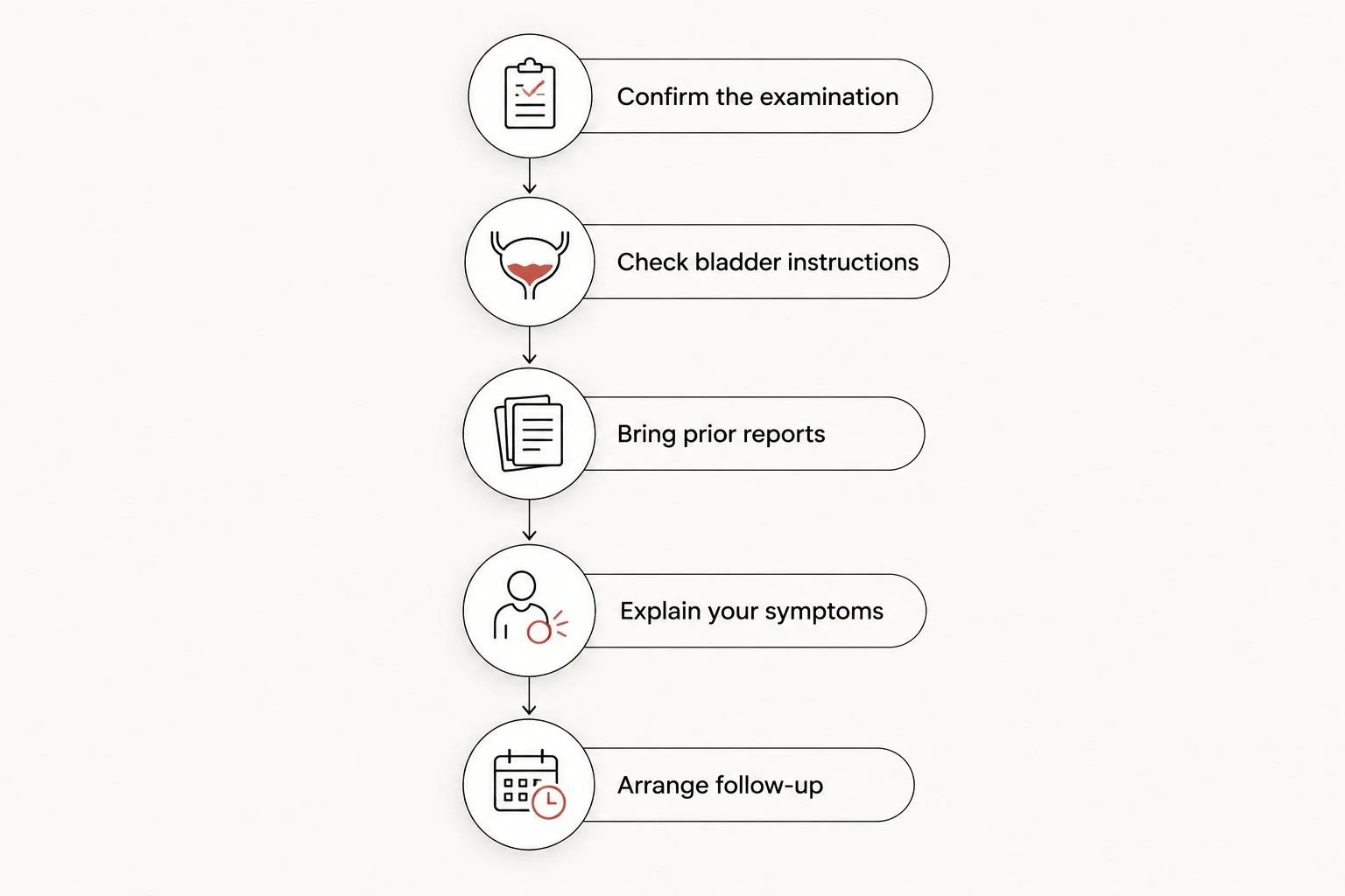
Preparation includes knowing who will review your results, not just how to fill your bladder.
If you are considering local private imaging, The Ultrasound Concierge is best for Keller and Willow Park patients seeking a private diagnostic ultrasound setting. Confirm that the requested pelvic examination and reporting arrangements meet your clinician’s needs; a calm appointment does not replace medical follow-up.
Use the contact page to ask about the requested examination and preparation before booking. Avoid assuming that every ultrasound appointment includes a fibroid assessment.
What happens after fibroids appear on ultrasound?
Finding fibroids does not automatically mean you need a procedure. Your clinician considers your symptoms, bleeding pattern, pregnancy plans, examination findings, and imaging report together.
If bleeding is heavy, your clinician can assess whether you also need testing for anemia or other causes of abnormal bleeding. A fibroid on the scan should not end that discussion if the symptoms suggest another problem.
If imaging does not clearly show the uterine cavity, saline infusion sonohysterography or hysteroscopy can help answer a more focused question. MRI is another option when anatomy remains unclear or detailed mapping is needed before treatment.
For a 2026 follow-up visit, bring the actual report and previous imaging when available. Ask: “Does this finding explain my symptoms, and what should happen if the symptoms continue?”
Can ultrasound miss a uterine fibroid?
Yes, ultrasound can miss a fibroid, particularly when it is small or the examination is technically limited. Persistent symptoms deserve follow-up even when no fibroid is reported; your clinician can decide whether to repeat imaging or investigate another cause.
Can an ultrasound tell whether a fibroid is cancerous?
No, ultrasound cannot definitively determine whether a uterine mass is cancerous. Most uterine fibroids are benign, but an unusual finding or concerning clinical history requires further evaluation rather than reassurance based on ultrasound alone.
Can I have fibroids without symptoms?
Yes, fibroids can be present without causing noticeable symptoms. An incidental finding does not automatically require treatment, but your clinician should explain whether observation or additional evaluation fits your situation.
When you should seek care promptly
Do not wait for a routine imaging appointment if you have sudden severe pelvic pain, fainting, or heavy bleeding accompanied by dizziness, weakness, chest discomfort, or shortness of breath. These symptoms need urgent medical assessment.
Pain or bleeding with a possible pregnancy also needs prompt evaluation. Do not assume a known fibroid explains a new symptom.
Bleeding after menopause requires medical evaluation even if you previously had fibroids. A familiar diagnosis should not become the reason a new concern goes unchecked.
FAQ
Can an ultrasound detect uterine fibroids if I have no symptoms?
Yes, ultrasound can detect uterine fibroids even when you have no symptoms. Fibroids are sometimes found during imaging requested for another reason, and the finding does not automatically mean treatment is needed.
Is an internal ultrasound better for finding fibroids?
Transvaginal ultrasound often provides more detailed views of the uterus, but it is not better for every imaging question. A transabdominal examination adds a wider view, and the approaches can complement each other.
Do I need a full bladder for a fibroid ultrasound?
You often need a full bladder for the transabdominal portion and an empty bladder for the transvaginal portion. Follow the imaging center’s specific instructions rather than a generic preparation schedule.
Can I have a pelvic ultrasound while I am on my period?
Menstruation does not usually prevent a diagnostic pelvic ultrasound. Tell the scheduling team about your bleeding and follow any instructions specific to the examination requested.
Can ultrasound tell the difference between fibroids and ovarian cysts?
Ultrasound often distinguishes a uterine fibroid from an ovarian cyst by showing where the finding originates and what it looks like. Some masses remain unclear and require additional evaluation.
Does a normal ultrasound rule out every cause of pelvic pain?
No, a normal ultrasound does not rule out every cause of pelvic pain. Persistent or worsening symptoms need clinical follow-up, including consideration of conditions that ultrasound does not reliably exclude.
What should I ask before booking private fibroid imaging in 2026?
Ask whether the appointment is a diagnostic pelvic examination appropriate for suspected fibroids, what preparation is required, and how results reach your clinician. The Ultrasound Concierge provides private diagnostic ultrasound services in Keller and Willow Park; confirm the requested examination before scheduling.
One last thing
The most useful question is not simply whether a fibroid is present. It is whether the finding changes your care.
Before leaving your 2026 follow-up appointment, ask your clinician to identify the fibroid’s location, explain whether the uterine cavity is affected, and outline what symptoms should trigger reassessment. A clear follow-up plan matters more than an image without an explanation.