
A routine fertility ultrasound cannot reliably confirm whether your fallopian tubes are blocked or open, although it can reveal abnormalities such as a fluid-filled, swollen tube. An ultrasound-based tubal patency test, such as hysterosalpingo-contrast sonography (HyCoSy), uses contrast to assess passage through the tubes; your clinician might instead recommend an X-ray test called hysterosalpingography (HSG).
TL;DR
For fertility ultrasound blocked fallopian tubes questions, routine scans cannot establish tubal patency.
HyCoSy uses ultrasound contrast; HSG uses X-rays and contrast to assess whether fallopian tubes are open.
A normal pelvic ultrasound does not rule out blocked tubes or every cause of infertility.
The Ultrasound Concierge offers private fertility ultrasound imaging; confirm the exact examination your clinician needs before booking.
Why this matters: A normal scan is reassuring about the structures examined, but it does not answer every fertility question. For your 2026 evaluation, ask whether the requested examination assesses your ovaries and uterus, tubal patency, or both.
The Ultrasound Concierge provides private diagnostic and fertility ultrasound services in Keller and Willow Park, Texas. The distinction between routine imaging and a dedicated tube test matters more than the appointment's general label.
Can a fertility ultrasound check for blocked fallopian tubes?
A standard fertility ultrasound can identify clues to tubal disease, but a dedicated patency test is needed to assess whether fluid passes through the tubes. Healthy fallopian tubes are usually difficult to see on routine ultrasound. Not seeing them is not evidence that they are blocked.
These examinations answer different questions:
Routine ultrasound
Best for: Assessing the uterus, ovaries, and visible pelvic abnormalities
Main benefit: Evaluates follicles, uterine lining, and findings such as cysts
Main limitation: Does not reliably establish whether tubes are open
HyCoSy
Best for: Ultrasound-based tubal patency assessment
Main benefit: Tracks contrast passage without X-rays
Main limitation: Requires a dedicated procedure; spasm or technical factors can affect interpretation
HSG
Best for: X-ray assessment of tubal patency
Main benefit: Shows the uterine cavity and contrast passage through the tubes
Main limitation: Uses ionizing radiation and contrast; apparent blockage sometimes needs clarification
Laparoscopy
Best for: Selected patients needing surgical pelvic assessment
Main benefit: Allows direct inspection of pelvic structures and dye passage
Main limitation: Requires surgery and anesthesia; not a routine first test for everyone
A report describing normal ovaries and a normal uterus does not automatically include a tubal assessment. Look for an explicit statement about the tubes and the method used to assess them.
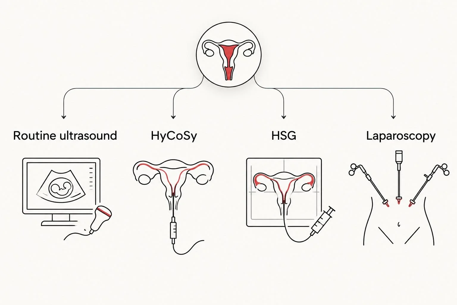
Routine pelvic imaging and dedicated tubal patency testing answer different questions.
Routine ultrasound: useful clues, not proof of open tubes
A transvaginal ultrasound gives a close view of the uterus and ovaries through a probe placed in the vagina, with your consent. It can assess developing follicles, the uterine lining, ovarian cysts, and some structural abnormalities.
A hydrosalpinx is a fallopian tube that has become swollen with fluid, usually because of obstruction. Ultrasound can sometimes identify its characteristic appearance. That finding warrants clinical review, but the absence of a hydrosalpinx does not rule out a blockage elsewhere.
Routine imaging also cannot show whether the microscopic structures inside a tube transport an egg normally. An open tube and a normally functioning tube are not the same finding. Your clinician interprets imaging alongside your history and the rest of your fertility evaluation.
HyCoSy: ultrasound with a dedicated contrast procedure
HyCoSy assesses tubal patency by introducing contrast through a small catheter passed through the cervix while ultrasound follows its movement. Passage through a tube and spill into the pelvis support that the tube is open.
Its advantage is that it uses ultrasound rather than X-rays. Its limitations include discomfort during catheter placement or fluid injection, technical difficulty, and temporary tubal spasm that complicates interpretation. Infection is a potential procedural risk, so your team should review relevant symptoms and history beforehand.
Do not assume that an appointment called a saline ultrasound includes HyCoSy. A standard saline-infusion sonohysterogram primarily examines the uterine cavity; the protocol must specifically address tubal patency to answer the blockage question.
HSG: contrast imaging with X-rays
HSG uses a catheter to introduce contrast into the uterus while X-rays record its path through the uterine cavity and fallopian tubes. Your clinician looks for filling of the tubes and contrast spilling from their ends.
HSG is an established option for evaluating suspected tubal blockage. It also has limitations: cramping can occur, the procedure uses radiation, and a result suggesting obstruction near the uterus can reflect spasm rather than a fixed blockage.
Tell the team about possible pregnancy, infection symptoms, and any previous reaction to contrast. An HSG result suggesting blockage needs interpretation, not an automatic conclusion that pregnancy is impossible. The location and extent of the finding influence the next step.
Laparoscopy: a surgical option for selected situations
During laparoscopy, a surgeon examines the pelvis through small abdominal incisions. Dye introduced through the cervix can show whether it passes through the fallopian tubes, while direct inspection can identify conditions such as adhesions or endometriosis.
The benefit is the ability to examine the outside of the tubes and surrounding pelvic structures. The drawback is the burden and risk of surgery and anesthesia. Laparoscopy is not a routine first-line tubal test for every person having difficulty conceiving.
Your clinician decides whether a surgical examination is justified by symptoms, history, other test results, or a separate reason for surgery. A routine ultrasound finding alone does not determine that decision.
Why tubal test results vary
A test result reflects both your anatomy and the conditions during the examination. The following factors help explain why apparently conflicting findings require clinical review:
The examination's purpose: Follicle monitoring and tubal patency testing use different methods and cannot substitute for each other.
The location of a blockage: Obstruction near the uterus differs from damage near the tube's outer end.
Temporary spasm: A tube can contract during contrast injection and appear blocked.
Technical conditions: Catheter position, contrast passage, and image quality influence what the examiner can establish.
Underlying pelvic disease: Prior inflammation, adhesions, or endometriosis can affect fertility beyond whether a tube appears open.
An inconclusive result is not a confirmed diagnosis. Ask what was demonstrated, what remains uncertain, and whether another examination would change your treatment plan.
Can I have blocked tubes even if my fertility scan is normal?
Yes, blocked fallopian tubes can be present despite a normal routine fertility ultrasound. A routine scan does not directly test passage through the tubes, and many blockages do not produce an obvious ultrasound abnormality.
The same distinction applies to regular periods and evidence of ovulation. Those findings address other parts of reproduction; they do not prove that an egg and sperm can travel through the tubes.
Does an open tube mean I can definitely get pregnant?
No, an open fallopian tube does not guarantee pregnancy. Conception also depends on ovulation, sperm factors, egg-related factors, the uterine environment, and other aspects of reproductive health.
If a test shows that only one tube is open, natural conception can still be possible. Your clinician should explain the result in the context of your age, history, and other findings rather than treating a single image as a prediction.
How should I prepare for a tubal patency appointment?
Before a 2026 appointment, get instructions for the specific examination rather than using general ultrasound preparation advice. A routine pelvic scan and a catheter-based tubal test do not have identical preparation requirements.
Confirm the exact test. Ask whether the order is for routine pelvic ultrasound, HyCoSy, HSG, or another procedure.
Check cycle timing. Tubal testing is commonly arranged after menstrual bleeding ends and before ovulation; follow your clinician's instructions about pregnancy prevention and testing.
Share relevant history. Mention pelvic infection, previous ectopic pregnancy, pelvic surgery, endometriosis, and contrast reactions.
Ask about comfort measures. Discuss pain relief, what sensations to expect, and how to request a pause.
Clarify follow-up. Find out who interprets the examination and who will explain its implications for your care.
Do not assume you need a full bladder, fasting, or a particular medication. Follow the performing team's instructions, especially if they differ from advice for an earlier scan.
Why does a history of chlamydia matter?
The link between chlamydia and infertility involves the risk of pelvic inflammatory disease and damage to the fallopian tubes. Chlamydia can occur without noticeable symptoms, and a previous infection is relevant even if you feel well now.
A history of chlamydia does not prove your tubes are blocked. It gives your clinician a reason to consider tubal factors when choosing tests. Treating an active infection is important, but treatment does not establish whether prior scarring exists or whether a tube is open.
Tell your clinician about previous infections without worrying that the information is irrelevant or embarrassing. It helps the team select an appropriate examination and assess precautions before a procedure involving the cervix and uterus.
When should I ask for a fertility evaluation?
If you are planning pregnancy in 2026, the timing of an evaluation depends on age and medical history. American Society for Reproductive Medicine guidance recommends evaluation after 12 months of trying to conceive for people younger than 35 years, or after 6 months for those aged 35 years or older.
Do not wait for those intervals if you have a known condition associated with infertility. A history of tubal disease, pelvic inflammatory disease, irregular or absent periods, or other relevant concerns warrants an earlier conversation with your clinician.
These thresholds guide when to seek evaluation; they are not instructions to order a tube test yourself. A fertility assessment also considers ovulation and, when relevant, semen analysis so that care does not focus on the tubes alone.
What should I ask a local ultrasound provider?
For a 2026 fertility ultrasound appointment, start with the clinical question on your order: are you monitoring follicles, examining the uterine lining, or investigating suspected tubal blockage? Ask whether the proposed examination can answer that question and whether a separate facility or specialist is needed.
The Ultrasound Concierge is best for Keller and Willow Park patients seeking private fertility ultrasound imaging, not a replacement for specialist tubal evaluation. Its concierge setting fits patients seeking a calm, personalized imaging visit; the examination's scope still determines what the visit can establish.
Before arranging care, contact the team to confirm the requested examination, preparation instructions, and how your referring clinician will receive the findings. Do not assume that general fertility imaging includes contrast-based tubal testing.
Keep the written report and bring it to your follow-up appointment. The most useful next step is a clear explanation of how the findings affect your care, not simply another scan without a defined question.
FAQ
Can a regular transvaginal ultrasound show blocked fallopian tubes?
A regular transvaginal ultrasound cannot reliably confirm blocked or open fallopian tubes. It can reveal findings such as a hydrosalpinx, but a normal scan does not exclude tubal blockage.
Is HyCoSy the same as a fertility ultrasound?
HyCoSy is a specialized ultrasound-based tubal patency procedure, not the same as routine fertility imaging. It introduces contrast through the cervix so the examiner can assess passage through the tubes.
Is HSG better than ultrasound for checking blocked tubes?
HSG is more appropriate than routine ultrasound for assessing tubal patency, but specialized contrast ultrasound is another option. Your clinician selects the examination based on your history and the clinical question.
Does saline ultrasound check whether both tubes are open?
A standard saline-infusion sonohysterogram does not necessarily establish whether both tubes are open. Ask whether the planned procedure specifically includes tubal patency assessment.
Will checking my fallopian tubes hurt?
Catheter-based tubal tests can cause cramping during catheter placement or contrast injection. Ask the performing team about pain relief and tell them if you need the procedure paused.
Can I get pregnant with one open fallopian tube?
Natural pregnancy can be possible with one open fallopian tube. Your individual outlook also depends on ovulation, sperm factors, the condition of the remaining tube, and other reproductive factors.
When should I seek urgent care after a tubal test?
Seek prompt medical advice for fever, worsening pelvic pain, foul-smelling discharge, or heavy bleeding after a tubal test. Severe pain, fainting, or breathing difficulty requires urgent medical assessment.
One last thing
For your 2026 care plan, pay attention to the difference between not seen, not assessed, and blocked. Those phrases do not mean the same thing, and none should be substituted for another when you discuss your report.
Bring one question to your follow-up: Did this examination actually test whether contrast passed through my fallopian tubes? That question separates reassuring pelvic images from an answer about tubal patency and helps you avoid leaving the appointment with false reassurance.