
Yes—having conceived through IVF does not, by itself, rule out a later 3D/4D ultrasound, but your fertility specialist or obstetric clinician should guide whether and when imaging is appropriate. Immediately after embryo transfer, follow your clinic’s pregnancy-testing and diagnostic ultrasound schedule; an elective scan cannot establish that implantation succeeded. Medical organizations discourage ultrasound performed solely for keepsake images, so discuss the purpose of any additional scan with your care team before booking.
TL;DR
A 3D 4D ultrasound after IVF embryo transfer does not replace fertility-clinic monitoring or prenatal diagnostic imaging.
ACOG discourages ultrasound solely for keepsake images; discuss additional pregnancy ultrasound with your obstetric clinician.
Early IVF ultrasound assesses pregnancy location and development, not detailed facial images.
Keep your clinic’s assigned due date and imaging schedule when arranging outside appointments.
Can you have a 3D/4D ultrasound after an IVF embryo transfer?
Yes, but distinguish an ultrasound needed for your care from an appointment intended mainly to create pictures. IVF conception alone does not determine the imaging method; the clinical question and stage of pregnancy do.
If you are arranging outside imaging in 2026, start with coordinating ultrasound monitoring with your IVF clinic. An outside appointment should support your clinic’s plan, not create a competing schedule.
Early diagnostic 2D ultrasound
Best for: Assessing pregnancy location and early development
Main benefit: Answers specific early-pregnancy questions
Main limitation: Does not produce detailed facial portraits
Medically indicated 3D ultrasound
Best for: Additional views for selected clinical questions
Main benefit: Reconstructs a volume into three-dimensional views
Main limitation: Does not replace a complete diagnostic examination
Medically indicated 4D ultrasound
Best for: Selected situations requiring moving three-dimensional views
Main benefit: Shows reconstructed images in motion
Main limitation: Is not necessary for every pregnancy assessment
Elective 3D/4D imaging
Best for: Personal viewing rather than medical assessment
Main benefit: Provides another way to see the pregnancy
Main limitation: Keepsake-only scanning is discouraged by medical organizations
These are different purposes, not a ladder from basic care to better care. A standard diagnostic examination remains valuable even when it does not produce a recognizable face.
Why this matters after IVF
After fertility treatment, waiting for the next appointment can feel especially difficult. Wanting another look is understandable, but an image and a medical assessment are not interchangeable.
Early pregnancy care asks whether the pregnancy is in the uterus, what structures are visible, and whether development matches the expected stage. Later prenatal care addresses anatomy, growth, and other clinical questions. Facial images answer a different kind of question.
For your 2026 care plan, decide which question you need answered before choosing an appointment. If your concern is whether the pregnancy is progressing, contact your treating team rather than relying on an elective viewing session.
Early 2D ultrasound: your clinic sets the timing
An early diagnostic ultrasound after IVF often occurs around 6–7 weeks of pregnancy, although your clinic sets the timing according to your treatment and clinical circumstances. Transvaginal imaging is commonly used because it provides useful views early in pregnancy.
That appointment is not scheduled a fixed number of weeks after every transfer. Pregnancy dating also accounts for the embryo’s age at transfer, so the transfer date is not the beginning of gestational age.
For example, on the day of a day-5 embryo transfer, obstetric gestational age is 2 weeks and 5 days. Use the dates supplied by your fertility clinic rather than restarting the pregnancy clock on transfer day.
An early scan also has limits. When findings are uncertain, your clinician interprets the images alongside your dates, symptoms, and blood-test results and determines whether follow-up is needed. Switching to a more visually detailed imaging mode does not remove those limits.
3D ultrasound: additional views, not earlier confirmation
A 3D ultrasound reconstructs ultrasound data into a three-dimensional image. Clinicians use it for selected diagnostic purposes, but a surface image is not a substitute for a complete examination.
Immediately after embryo transfer, there is no developed fetal face to photograph. Buying a different imaging format does not make implantation visible sooner or establish that the pregnancy will continue normally.
Later in pregnancy, recognizable facial views depend on fetal development, position, and the available acoustic window. Hands, the placenta, or surrounding structures can obscure the face. An unclear portrait does not, by itself, establish a medical problem.
4D ultrasound: movement does not mean a fuller assessment
A 4D ultrasound displays three-dimensional images over time, creating a moving view. Its distinction from a still 3D image is motion, not an automatic increase in diagnostic certainty.
A moving facial image does not establish that all organs are developing normally. It also does not replace the routine anatomy examination, commonly performed at 18–22 weeks of pregnancy under your prenatal clinician’s direction.
For appointments in 2026, ask whether the proposed examination is diagnostic, elective, or includes both purposes. Do not infer the scope from the words 3D or 4D alone.
Why timing and image quality vary
There is no single post-transfer date that answers every patient’s imaging needs. Your clinician and imaging provider need to consider the purpose of the examination as well as the pregnancy stage.
Gestational age: Early development and later facial imaging require different expectations. Your fertility clinic’s dates provide the starting point.
Clinical purpose: Confirming pregnancy location is different from assessing anatomy or obtaining a facial view. The examination should match the question.
Symptoms and findings: Pain, bleeding, or an unresolved earlier result require clinical direction rather than a routine elective booking.
Fetal position: A baby facing away from the probe or covering the face can limit surface images.
Surrounding structures and fluid: The placenta, uterine wall, and fluid around the face affect the view available to the sonographer.
Multiple pregnancy: Twins introduce additional positioning and imaging challenges, while their medical monitoring follows the treating clinician’s plan.
These factors explain why a particular image cannot be promised. They are not reasons to extend scanning indefinitely or arrange repeated sessions without discussing their purpose.
How to coordinate an outside ultrasound after IVF
Before scheduling an additional appointment in 2026, confirm the clinical plan and the information the outside provider needs. A short conversation with your fertility or obstetric team helps keep the appointment’s purpose clear.
Confirm your dates. Obtain your assigned due date and current gestational age from your fertility clinic. Include the transfer date and embryo age when requested.
Clarify the purpose. Explain whether you need an ordered diagnostic examination, have a symptom, or are asking about personal viewing. These are not interchangeable requests.
Ask your clinician. Discuss whether additional imaging is appropriate and whether any restrictions or unresolved findings affect the decision. Do not change medication or monitoring instructions yourself.
Check the examination. Ask who performs the scan, whether a diagnostic report is included, and who interprets medical findings. A photograph alone is not a report.
Plan follow-up. Identify who receives the report and whom you should contact with questions. Keep your existing prenatal appointments unless your treating clinician changes them.
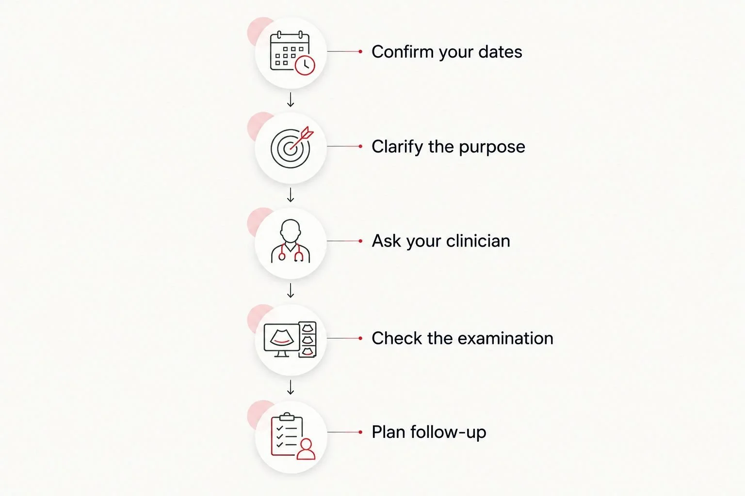
Agree on the purpose and follow-up before arranging outside imaging.
The Ultrasound Concierge provides private diagnostic and elective pregnancy ultrasound services in the Keller and Willow Park, Texas area. The Ultrasound Concierge is best for local patients seeking a private ultrasound setting. That setting does not change which examination your clinician recommends or what an elective image can establish.
Review The Ultrasound Concierge’s ultrasound services with your appointment’s purpose in mind. Ask about the specific examination and reporting arrangements rather than assuming that every pregnancy session provides the same assessment.
Is ultrasound safe after an embryo transfer?
Ultrasound uses sound waves rather than ionizing radiation. However, that distinction is not a reason to treat unlimited scanning as necessary or risk-free.
The American College of Obstetricians and Gynecologists discourages ultrasound performed solely for keepsake images. The U.S. Food and Drug Administration also discourages nonmedical fetal imaging, and the American Institute of Ultrasound in Medicine supports prudent use by appropriately trained professionals with exposure kept as low as reasonably achievable.
For a 2026 decision, the practical recommendation is to follow medically indicated imaging and discuss any additional appointment with your clinician. A private setting, a short appointment, or the label 4D does not replace professional judgment about whether the scan is appropriate.
Can a 3D/4D scan replace my IVF clinic’s first ultrasound?
No—a 3D/4D viewing session cannot replace the diagnostic assessment your fertility clinic has scheduled. An early examination evaluates questions such as pregnancy location and visible development, with findings interpreted in clinical context.
Even if an outside provider offers diagnostic imaging, your fertility clinic must determine whether that particular examination meets its needs. Do not cancel clinic monitoring because another appointment includes pictures or video.
Do I need to wait until after my anatomy scan?
There is no universal rule requiring every IVF patient to schedule additional imaging after the anatomy scan. Your clinician determines the appropriate sequence, and an elective session does not replace the routine 18–22 weeks anatomy examination.
If your anatomy examination identifies a concern or requires additional views, complete the recommended diagnostic follow-up. A clearer facial portrait is not an alternative to evaluating the unresolved finding.
What if I have pain or bleeding before the appointment?
Contact your fertility or obstetric team promptly about pain or bleeding after IVF. Do not wait for an elective session to assess symptoms.
Severe abdominal or pelvic pain, fainting, shoulder-tip pain, or heavy bleeding warrants urgent medical assessment. IVF does not eliminate the possibility of an ectopic pregnancy, and an elective facial-imaging appointment is not the appropriate setting to evaluate an emergency.
If you cannot reach your team and symptoms are severe, seek emergency care. A previously reassuring image does not override a new symptom.
FAQ
Can I have a 3D 4D ultrasound after IVF embryo transfer?
IVF conception alone does not rule out later 3D/4D imaging, but your fertility or obstetric clinician should guide whether additional imaging is appropriate. Follow your clinic’s early diagnostic schedule, and remember that medical organizations discourage keepsake-only ultrasound.
Can a 3D ultrasound show implantation after my transfer?
A 3D ultrasound cannot establish successful implantation immediately after transfer. Your fertility clinic uses its pregnancy-testing schedule and appropriately timed diagnostic ultrasound to assess the pregnancy.
When is the first ultrasound after IVF usually done?
The first diagnostic ultrasound after IVF often occurs around 6–7 weeks of pregnancy, with timing set by your fertility clinic. Symptoms, test results, and individual circumstances affect the plan.
Does a transvaginal ultrasound dislodge an embryo?
A properly performed transvaginal ultrasound does not enter the uterine cavity or dislodge an implanted embryo. The probe remains in the vagina; follow your clinic’s instructions about the examination.
Is 4D ultrasound better than 2D for checking an early IVF pregnancy?
4D ultrasound is not automatically better than 2D ultrasound for assessing an early IVF pregnancy. Standard diagnostic imaging answers early clinical questions, while 4D adds moving three-dimensional views.
Can a private ultrasound replace my anatomy scan?
An elective private ultrasound cannot replace your diagnostic anatomy examination. The routine anatomy examination commonly occurs at 18–22 weeks of pregnancy and evaluates structures beyond the baby’s face.
What should I tell The Ultrasound Concierge before an appointment?
Tell The Ultrasound Concierge your clinic-assigned gestational age, the purpose of the appointment, and any relevant clinician instructions. Discuss symptoms or unresolved medical findings with your treating team before arranging elective imaging.
One last thing
A more recognizable picture is not necessarily a more informative medical examination. After IVF, the useful next scan is the one that answers the right question at the right stage—not automatically the one with the most dimensions.
Keep your fertility clinic’s dates and instructions together when planning appointments in 2026. If your goal is reassurance about development, ask your treating clinician what assessment answers that concern before choosing an imaging format.