
Yes. An ultrasound can detect low amniotic fluid by measuring fluid pockets around your baby, using the amniotic fluid index or the single deepest pocket method. The measurement estimates fluid volume rather than measuring every drop, and your prenatal clinician must interpret it alongside your pregnancy stage, symptoms, and other findings.
TL;DR
Can an ultrasound detect low amniotic fluid? Yes—diagnostic ultrasound estimates amniotic fluid using measured pockets.
An amniotic fluid index of 5 cm or less is a commonly used threshold for oligohydramnios.
A single deepest pocket below 2 cm is another established threshold for low amniotic fluid.
The Ultrasound Concierge offers private pregnancy ultrasound; suspected fluid leakage needs prompt assessment from your prenatal team.
The Ultrasound Concierge provides diagnostic and elective ultrasound imaging in Keller and Willow Park, Texas. When fluid levels are the concern, the appointment's medical purpose matters more than the type of keepsake images offered.
Can an ultrasound detect low amniotic fluid?
Ultrasound is the standard imaging method used to assess whether amniotic fluid is low. The medical term is oligohydramnios. During an abdominal scan, the sonographer identifies fluid pockets that do not contain the umbilical cord or fetal body parts and measures their vertical depth.
There are two established measurement methods. Both produce measurements in centimeters, but they are not interchangeable.
Amniotic fluid index, or AFI
What it measures: The sum of vertical fluid-pocket measurements from 4 uterine quadrants
Common low-fluid threshold: 5 cm or less
Best suited to: Assessments using an AFI-based protocol
Limitation: Identifies more pregnancies as low-fluid than the single-pocket method, which can increase interventions
Single deepest pocket, or SDP
What it measures: The vertical depth of the largest suitable fluid pocket
Common low-fluid threshold: Less than 2 cm
Best suited to: Fluid surveillance using a single-pocket protocol
Limitation: Describes one pocket rather than measuring total fluid volume
These thresholds are established obstetric criteria, not home-testing targets. A result needs clinical interpretation, particularly in twin pregnancies, where clinicians assess fluid in each amniotic sac rather than applying a whole-uterus AFI in the same way.
Ask which method appears on your report before comparing your measurement with a number you found online.
Why this matters in 2026
Low fluid is a finding that needs an explanation, not a diagnosis of one specific problem. It can occur with ruptured membranes, placental problems, certain fetal kidney or urinary tract conditions, or a pregnancy continuing beyond its due date.
It also does not automatically mean you need immediate delivery. Your clinician considers how far along you are, whether your membranes have ruptured, fetal growth, and evidence of your baby's well-being before recommending monitoring or delivery.
The practical distinction is straightforward: a reassuring picture of your baby's face does not answer the same question as a documented fluid assessment. Choose diagnostic evaluation when the concern is medical.
Single deepest pocket: less than 2 cm
A single deepest pocket below 2 cm is an established ultrasound criterion for oligohydramnios. Your report might call this the deepest vertical pocket, DVP, maximum vertical pocket, MVP, or SDP.
The sonographer looks for the largest suitable pocket and measures its vertical depth. The pocket must be free of fetal parts and umbilical cord; color Doppler can help identify cord within a suspected fluid pocket.
The American College of Obstetricians and Gynecologists uses a deepest vertical pocket below 2 cm in its guidance on isolated oligohydramnios and antenatal fetal surveillance. That threshold identifies a finding requiring clinical attention; it does not independently determine the cause or delivery plan.
A measurement close to the cutoff deserves context. Ask whether it was technically adequate, whether prior measurements are available, and whether other findings change the interpretation.
Amniotic fluid index: 5 cm or less
An AFI of 5 cm or less is another commonly used criterion for oligohydramnios. To calculate AFI, the examiner divides the uterus into 4 quadrants, measures a suitable vertical fluid pocket in each, and adds the measurements.
The total is a length-based estimate, not the amount of fluid in milliliters. An AFI of 5 cm therefore does not mean there are only 5 cm of fluid surrounding your baby everywhere.
AFI and the single deepest pocket method can classify the same pregnancy differently. Comparative evidence summarized in Cochrane reviews found that using AFI identifies more cases of oligohydramnios and leads to more inductions without improving several measured pregnancy outcomes compared with the single-pocket method.
A different classification does not mean someone performed the scan incorrectly. Your prenatal team should explain which method guides your care and why.
Why amniotic fluid measurements vary
A fluid measurement is a snapshot. The following factors help explain why measurements or their meaning differ between appointments:
Measurement method: AFI adds several pockets; SDP records the largest suitable pocket. Compare like with like.
Fetal position: Your baby's position changes which pockets are accessible for measurement.
Technique: Probe pressure, pocket selection, and identification of the umbilical cord affect the reading.
Pregnancy stage: Expected fluid patterns change as pregnancy progresses, particularly near and beyond the due date.
Membrane status: Leaking amniotic fluid can reduce fluid volume and requires assessment beyond the ultrasound measurement.
Maternal and fetal conditions: Placental function, fetal urine production, and certain medications can affect fluid volume.
A change between reports is a reason to ask questions, not to interpret the numbers on your own. Your clinician decides whether the difference represents a meaningful change, measurement variation, or a need for additional evaluation.
When reviewing a 2026 ultrasound report, keep the measurement method beside the result. A number separated from its label is easy to misunderstand.
What happens during a fluid assessment?
A fluid check generally uses an abdominal ultrasound probe and gel on your abdomen. The fluid measurements themselves are part of the scan, not a needle procedure.
The next steps depend on why the assessment was requested:
Clinical question: Tell the team about fluid leakage, changes in movement, your pregnancy stage, and the reason your clinician requested imaging.
Fluid measurement: The examiner records the requested AFI or deepest-pocket measurement.
Additional assessment: Depending on the clinical question, the examination can include fetal growth, movement, or other observations.
Clinical review: Your prenatal clinician interprets the report and explains the follow-up plan.
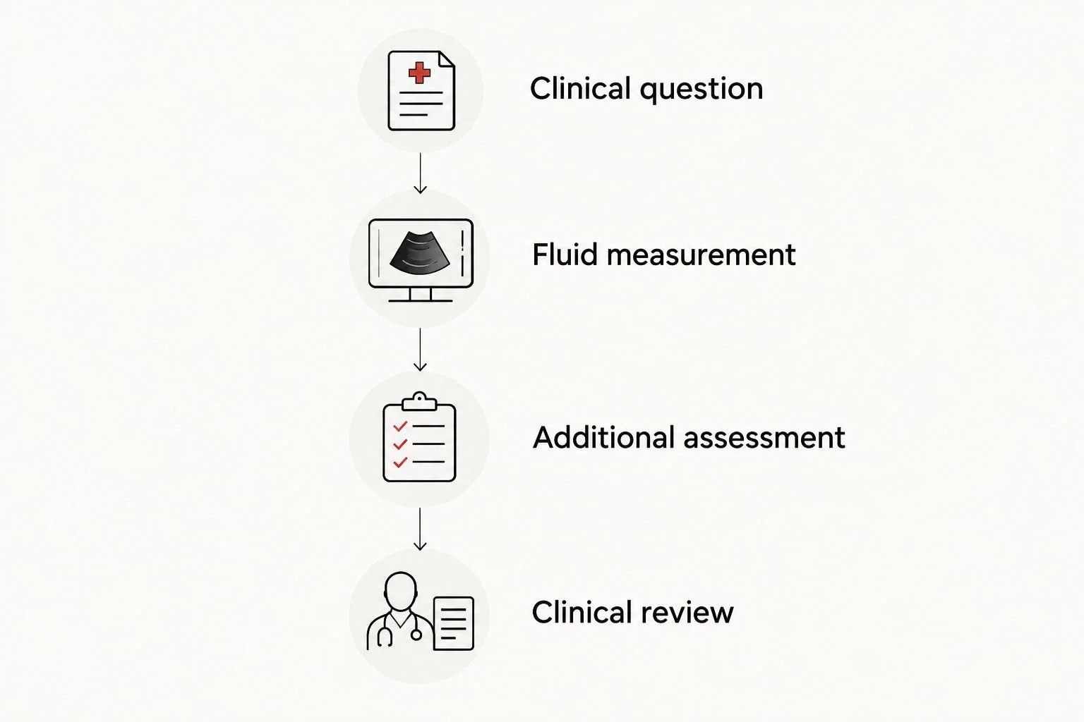
The fluid measurement is one part of an assessment that ends with a clinical plan.
Before leaving, ask who will review the findings and how you will receive instructions. Do not assume that receiving images means your prenatal clinician has received the report.
For a 2026 appointment, bring the imaging request and relevant previous reports if your care team asks for them. Follow the facility's preparation instructions rather than changing your fluid intake to try to influence the measurement.
Can ultrasound tell why the fluid is low?
Ultrasound can identify clues, but it does not establish every cause. It can assess fetal growth, examine visible kidney and bladder anatomy, and evaluate other findings that help your clinician narrow the possibilities.
Suspected membrane rupture needs a clinical assessment. A low fluid measurement can support that concern, but ultrasound alone cannot reliably confirm or exclude a leak.
Your clinician also reviews your health history, blood pressure, medications, and symptoms. Do not stop a prescribed medicine because you read that some medications affect amniotic fluid; ask the prescriber for guidance.
Sometimes the evaluation does not identify a clear cause. That does not make follow-up unnecessary: the measured fluid level and the rest of the pregnancy assessment still guide care.
Can a keepsake ultrasound check amniotic fluid?
An elective session is not a substitute for a clinician-directed diagnostic fluid assessment. The distinction is the examination's purpose, documented measurements, and clinical interpretation—not whether the equipment can produce detailed pictures.
Diagnostic obstetric ultrasound
Best for: A medical question such as suspected low fluid
Benefit: Documents measurements for clinical interpretation
Limitation for a fluid concern: Does not replace examination or testing for every possible cause
Elective keepsake ultrasound
Best for: Viewing and preserving pregnancy images
Benefit: Provides a non-diagnostic viewing experience
Limitation for a fluid concern: Does not replace a medical fluid assessment or prenatal surveillance
The Ultrasound Concierge is best for Keller and Willow Park patients seeking private ultrasound imaging, not a replacement for ongoing obstetric care. Its diagnostic and elective services serve different purposes; ask whether the proposed appointment addresses the medical question on your clinician's request.
A private setting offers a different visit experience, but it does not change the need for clinical follow-up. If you suspect leaking fluid or notice reduced movement, contact your prenatal team rather than booking an elective session for reassurance.
What should I do if my report says low fluid?
Contact the clinician managing your pregnancy for an interpretation and a specific plan. Ask whether the result meets the low-fluid threshold, whether your membranes need assessment, and whether additional monitoring is needed.
Monitoring can include repeat ultrasound, a nonstress test, or a biophysical profile. The timing depends on the overall clinical picture; there is no single repeat-scan interval appropriate for every pregnancy with low fluid.
Delivery decisions also depend on pregnancy stage and associated findings. A fluid measurement alone is not enough to decide whether induction is needed.
Before you finish the conversation, write down:
What the measurement was and which method was used.
Whether the finding is isolated or accompanied by another concern.
Where and when the next assessment should happen.
Which symptoms mean you should seek care sooner.
Keep that plan with your 2026 prenatal records so you do not have to reconstruct instructions from memory.
When should I call instead of waiting for a scan?
Contact your prenatal clinician or labor-and-delivery triage promptly for a gush or ongoing trickle of fluid, reduced fetal movement, vaginal bleeding, fever, or significant abdominal pain. Follow your maternity team's instructions about where to be evaluated.
You cannot reliably distinguish amniotic fluid from urine or discharge at home. Likewise, a normal fluid measurement from an earlier appointment does not rule out a new problem today.
Do not wait for an elective appointment to evaluate a possible fluid leak or reduced movement. If you cannot reach your prenatal team, seek urgent maternity assessment rather than waiting for a routine imaging opening.
FAQ
Can an ultrasound detect low amniotic fluid before I notice symptoms?
Yes. Ultrasound can detect low amniotic fluid even when you have not noticed symptoms. Symptoms alone cannot establish whether your fluid level is normal.
What ultrasound number means amniotic fluid is low?
An AFI of 5 cm or less or a single deepest pocket below 2 cm are established low-fluid criteria. Your clinician interprets the result using the measurement method and your pregnancy's circumstances.
Does low amniotic fluid mean my water has broken?
No. Low amniotic fluid has several possible causes, including membrane rupture. A suspected leak needs clinical assessment because ultrasound alone cannot confirm or exclude it.
Can drinking water fix low amniotic fluid?
Drinking water is not a reliable treatment for every cause of low amniotic fluid. Hydration can affect measurements in some circumstances, but it does not repair ruptured membranes or replace assessment of placental or fetal conditions.
Does low amniotic fluid always mean I need induction?
No. Delivery decisions depend on gestational age, membrane status, fetal well-being, and associated findings. Your prenatal clinician determines whether monitoring or delivery is appropriate.
Can a normal ultrasound rule out leaking amniotic fluid?
No. A normal fluid measurement does not reliably rule out ruptured membranes. Contact your prenatal team if you notice a gush or ongoing watery leakage.
Can I ask The Ultrasound Concierge about a fluid assessment in 2026?
Yes. The Ultrasound Concierge offers diagnostic pregnancy ultrasound, so you can ask whether an appointment matches your clinician's imaging request. Suspected fluid leakage or reduced movement requires prompt prenatal assessment rather than waiting for a routine appointment.
One last thing
A clear image is not a fluid measurement. Before accepting reassurance from a scan, ask for the documented AFI or deepest-pocket result and who will interpret it.
That simple question separates seeing your baby from answering the medical concern that brought you in. You deserve both an understandable explanation and a clear next step.